Encephalitis: Insights from Advanced Imaging and a 20-Year Archive
Encephalitis, the inflammation of the active tissues of the brain, can arise from infections or autoimmune responses where the body’s immune system mistakenly attacks the brain. The recognition of autoimmune causes has contributed to an increasing number of diagnosed cases. While mild cases often resolve completely, encephalitis remains a serious condition, with a mortality rate of approximately 10% despite advancements in diagnosis and treatment.
Magnetic Resonance Imaging (MRI) and Magnetic Resonance Spectroscopy (MRS) play a pivotal role in diagnosing and monitoring patients with infectious and inflammatory encephalitis. At our center, a 20-year archive of diverse cases has been curated, offering invaluable insights into the imaging findings of this complex condition. Below, we present three notable cases from this extensive archive.
Case 1: Herpes Simplex Virus Encephalitis (HSV-1)
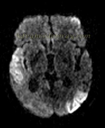
A 45-year-old male patient diagnosed with Herpes Encephalitis, presenting with bilateral temporal and parietal signal intensity abnormalities on MRI. Findings include cortical hemorrhage, microbleeding, and diffusion restriction, confirming a case of HSV-1 encephalitis.

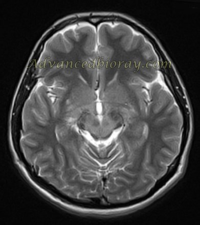
Case 2: Neuro-Behçet’s Disease
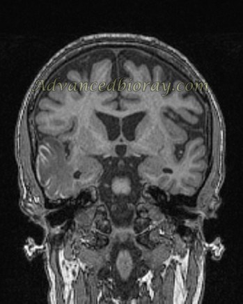
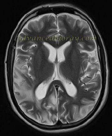
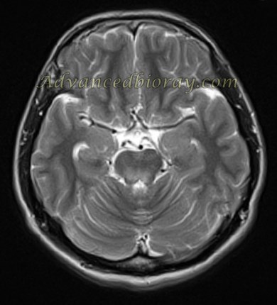
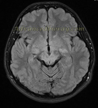
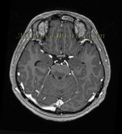
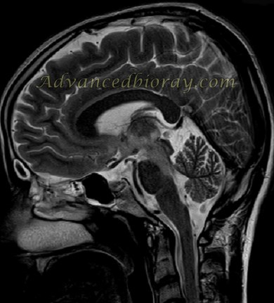
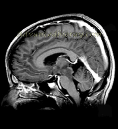
MRI Axial T2-weighted (T2W) sequences at the level of the basal ganglia and midbrain revealed hyperintense signals in both thalami, the posterior limb of the internal capsule, the posterior lentiform nucleus, and the midbrain. T2W coronal sequences showed hyperintense signals in both thalami, the midbrain, and bilateral pons, forming a characteristic “waterfall” appearance. These imaging findings supported the diagnosis of neuro-Behçet’s disease.

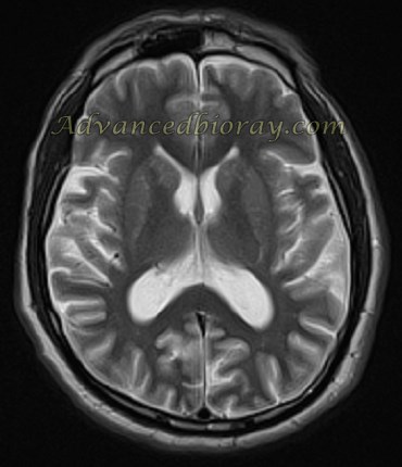
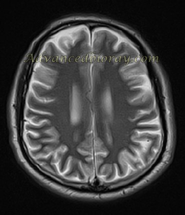
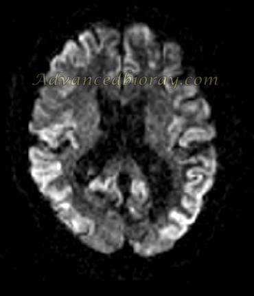
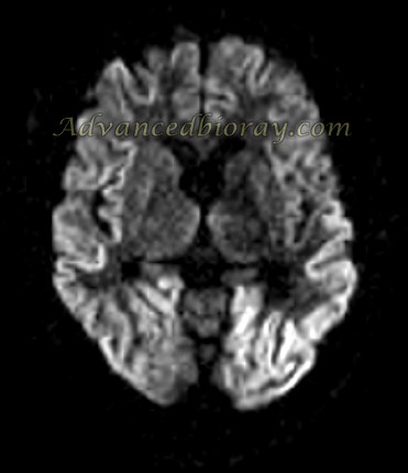
Case 3: Creutzfeldt-Jakob Disease (CJD)
A known case of Creutzfeldt-Jakob Disease (CJD) presenting with diffusion restriction in the cortex of both hemispheres on MRI.
Additional findings included mild cortical atrophy, characteristic of the disease.