Magnetic resonance imaging (MRI) has become an indispensable tool in the diagnosis of abdominal and pelvic disorders, serving as a complement to traditional imaging methods such as ultrasound and computed tomography (CT). Its role has significantly expanded in recent years, particularly for non-traumatic conditions and follow-up evaluations, owing to its high-resolution imaging and lack of ionizing radiation. These characteristics make MRI particularly advantageous for pregnant patients, where it can aid in diagnosing a variety of maternal abdominal and pelvic conditions as well as fetal anomalies.
MRI is most effective when used alongside other imaging modalities, allowing a comprehensive assessment while acknowledging its strengths and limitations. Despite its growing application in abdominal and pelvic imaging, the availability of MRI remains constrained by the number of scanners. However, its importance in the diagnosis and management of abdominal and pelvic disorders continues to rise.
Several noteworthy cases are presented below:
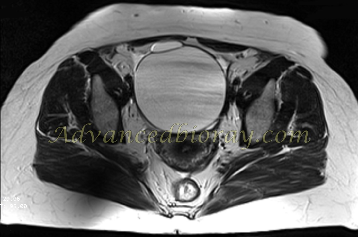
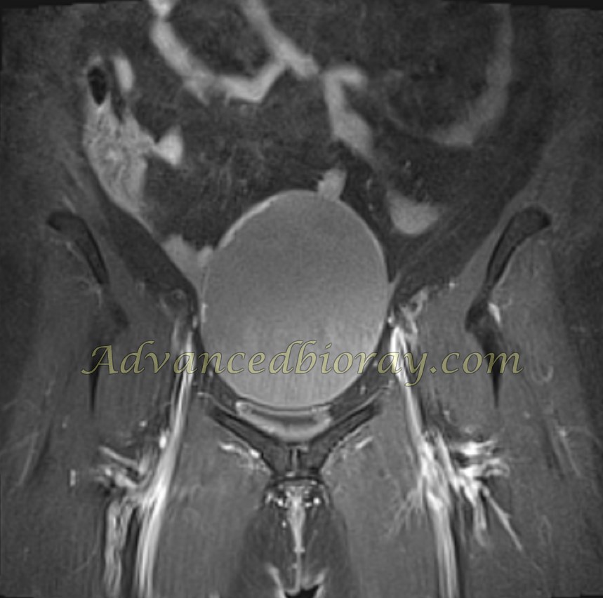
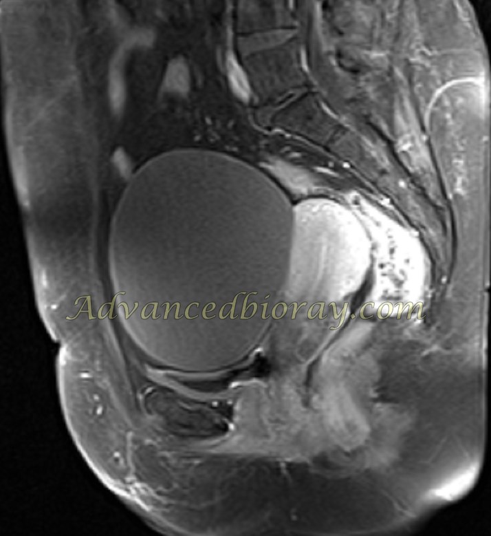
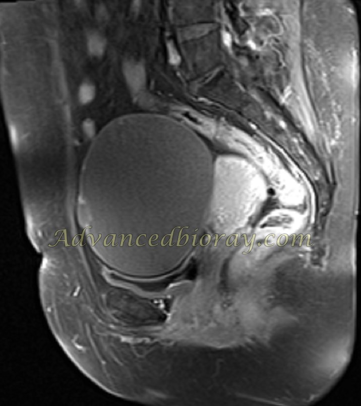
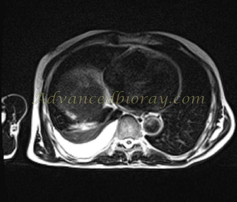
Case No 1 : 46-year-old woman with abdominal pain:
MRI with contrast revealed two cystic lesions in the ovaries. The left ovary contains a larger cyst measuring 88×85 mm, while the right ovary has a smaller cyst measuring 23×22 mm. The larger cyst demonstrates abnormal nodular enhancement along the inner wall, suggesting the possibility of cystadenomas.
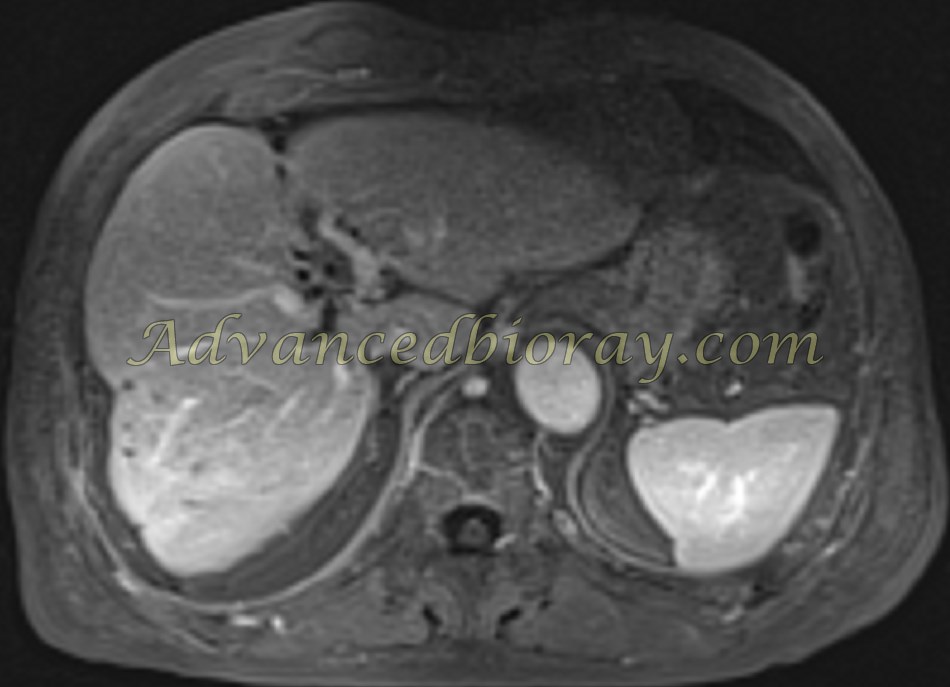
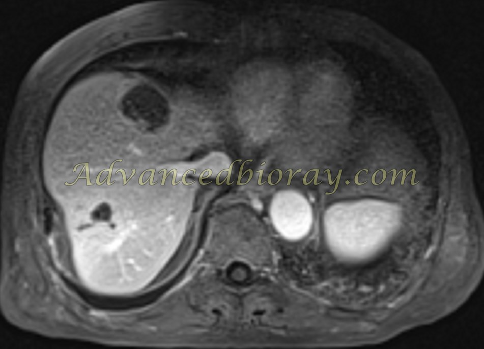
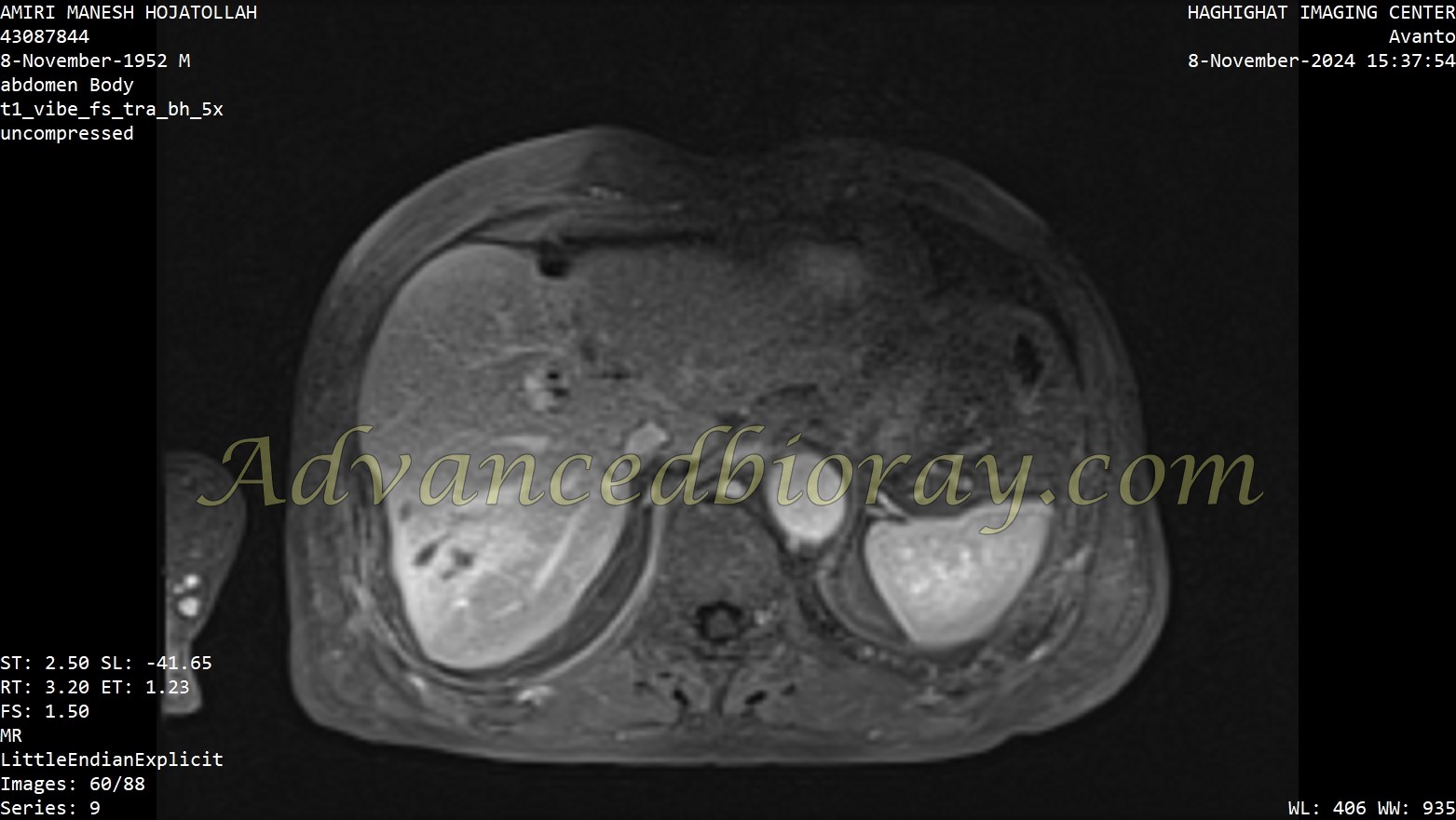
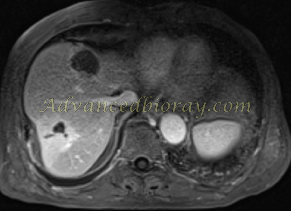
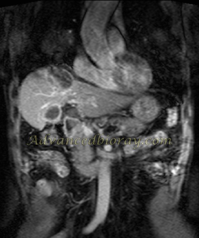
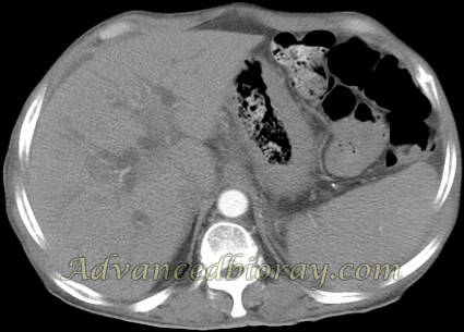
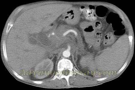
Case No 2 : 72-year-old man with a known history of HCC and prior TACE:
MRI shows a primary tumor in liver segment 8, measuring 37×35 mm, with a small enhancing component at the posterior wall, suggestive of a viable tumor component. Additionally, interparenchymal biloma is observed in the right liver lobe, along with evidence of gallbladder wall thickening. A moderate pleural effusion is noted in the right lung.
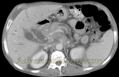
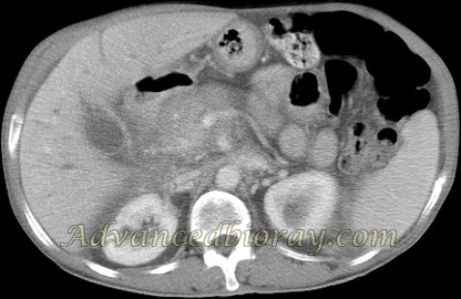
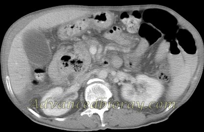
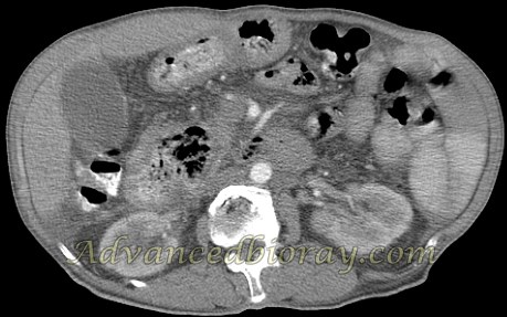
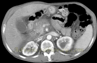
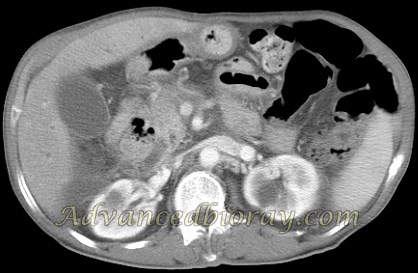
Case No 3 : 72-year-old woman with a known case of liver cirrhosis and portal hypertension:
CT reveals moderate to severe free fluid in the abdominal and pelvic cavities, accompanied by mild splenomegaly. Varicose veins are observed around the gastric fundus and the distal esophagus. There is diffuse intrahepatic bile duct (IHBD) dilation and common bile duct (CBD) dilation, measuring up to 18 mm.
An exophytic cystic lesion measuring 57×52 mm is noted in the D3 segment of the duodenum, containing food residue. This finding is suggestive of a large duodenal diverticulum.