Neuroimaging, particularly MRI, plays a pivotal role in diagnosing and characterizing spinal cord, spinal vertebral, and disk space lesions. It is indispensable in evaluating diskopathies and spondylotic changes, providing critical insights into herniated disk compression on nerve roots and the spectrum of spondylotic alterations. Additionally, MRI excels in localizing cord lesions and determining their etiology, which may include traumatic, inflammatory, infectious, compressive, or neoplastic conditions.
MRI’s ability to differentiate between extradural, intradural-extramedullary, and intramedullary lesions through specific imaging features aids in narrowing the differential diagnosis. It is also a key tool in evaluating primary spinal tumors, whether benign or malignant, by assessing their extent and relationships with adjacent structures.
In our archive, the high volume of daily spinal and cord MRI studies has enabled us to gather an extensive archive of interesting and educational cases, fostering knowledge expansion in this field and contributing to structured algorithmic approaches. Below, we present some illustrative cases:
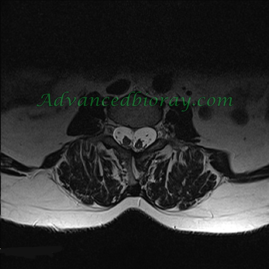
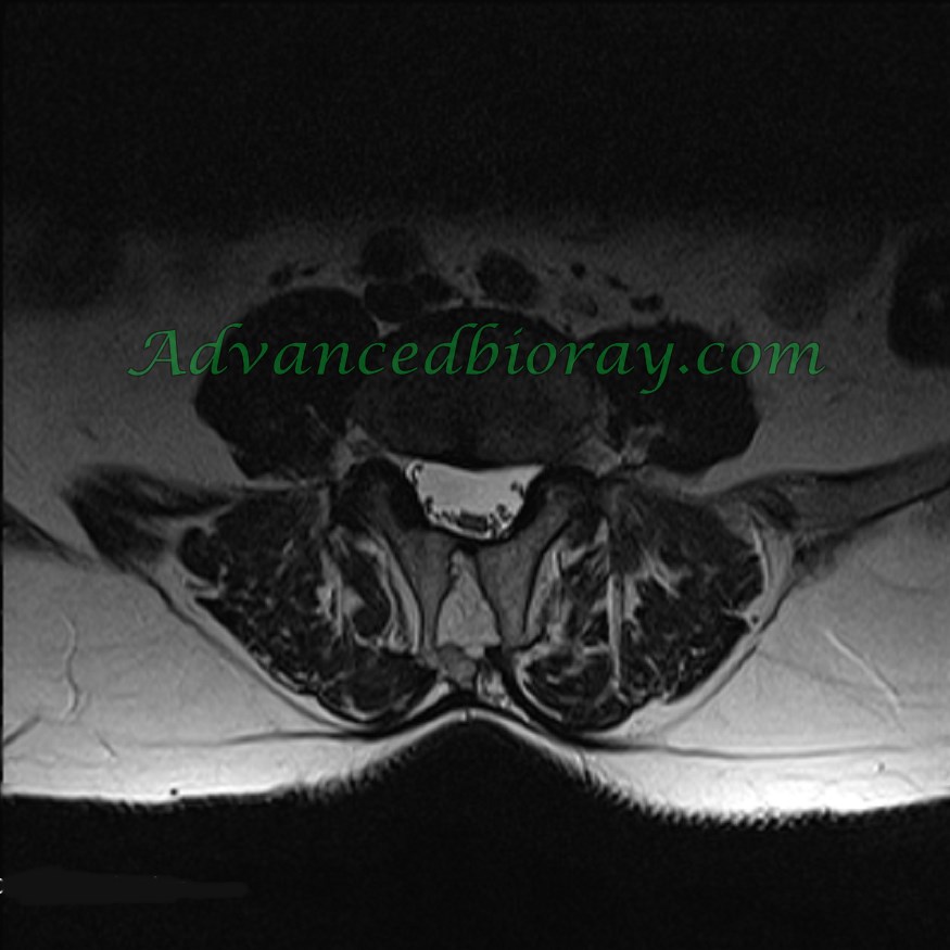
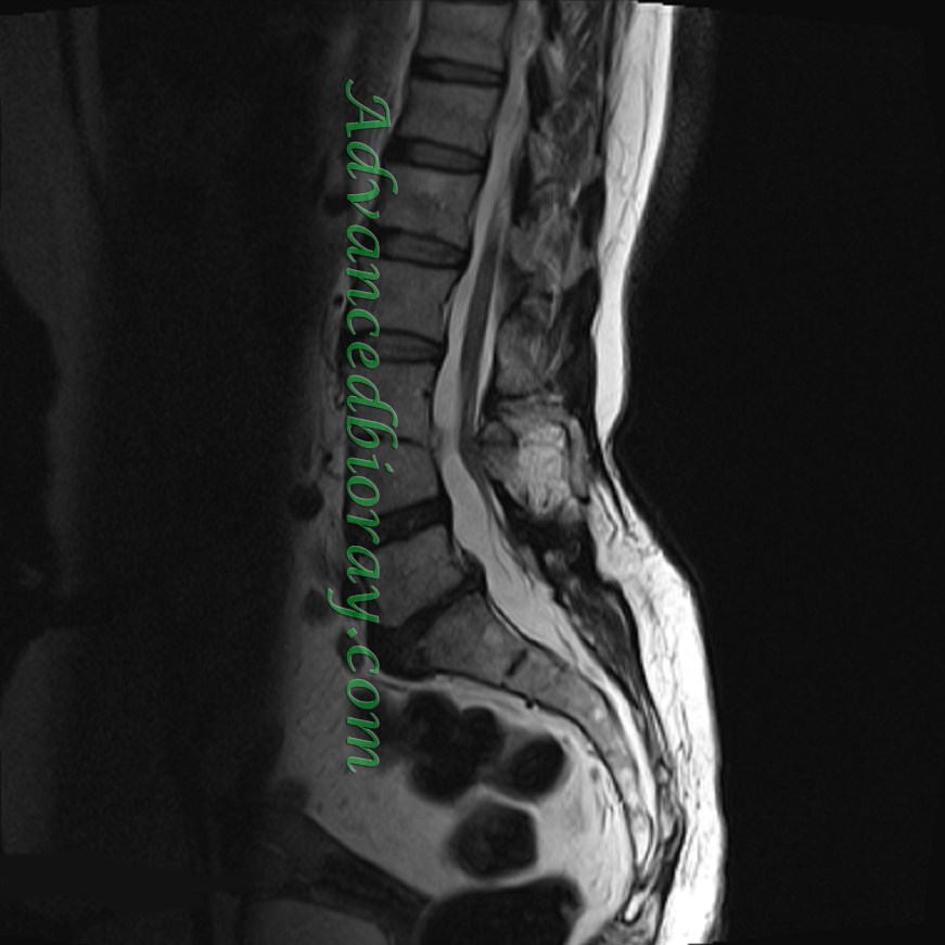
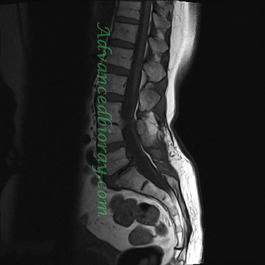
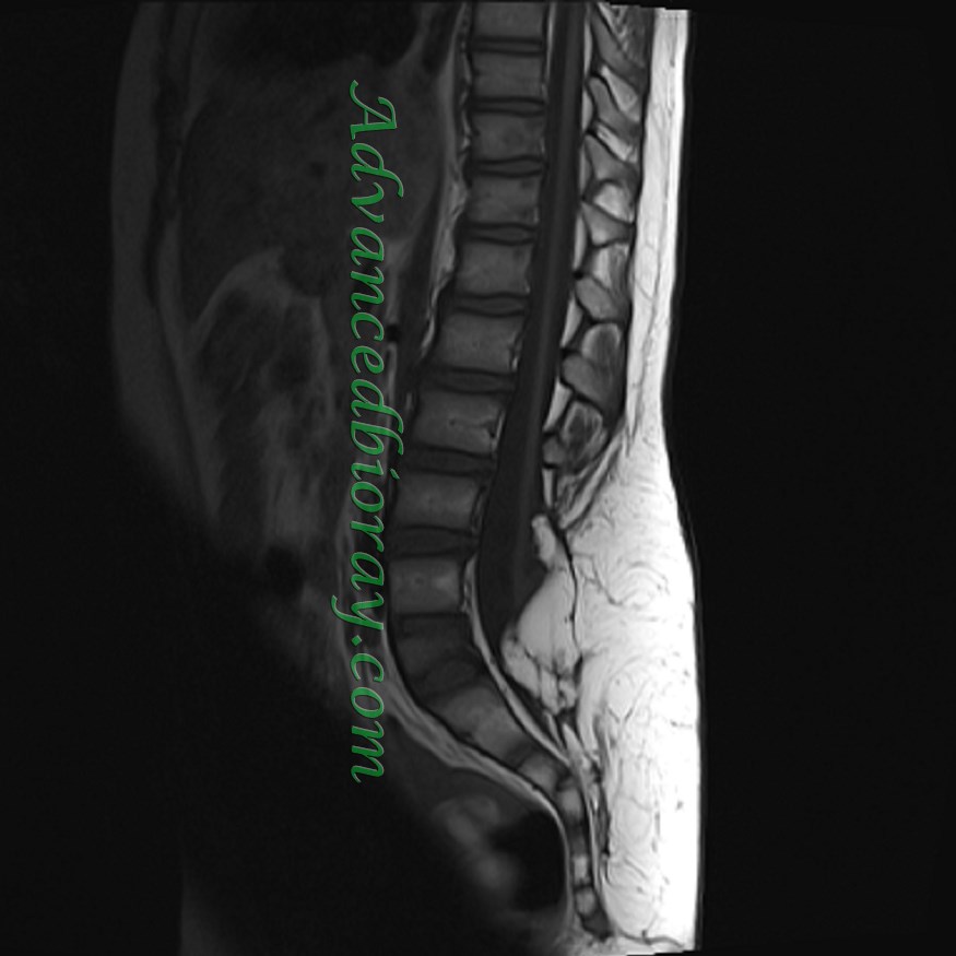
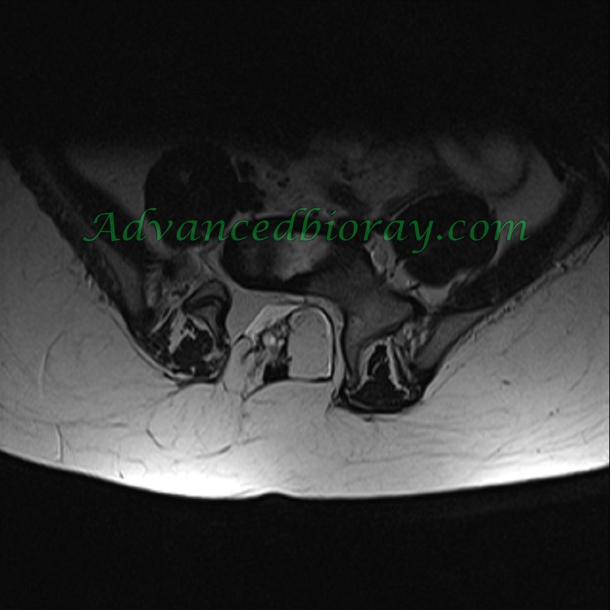
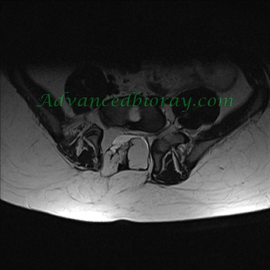
Case No 1 :
33-year-old man with complaints of low back pain (LBP).
MRI findings: Typical features of dysraphism, including a low-lying conus medullaris, vertebral fusion at L2-L3, diastematomyelia, and a bony septum.
Case No 2 :
10-year-old boy with spinal dysraphism.
MRI findings: Spina bifida with a typical lipomyelomeningocele, tethered cord terminating at the L5-S1 level, and distortion of the normal nerve root anatomy in the sacral canal.
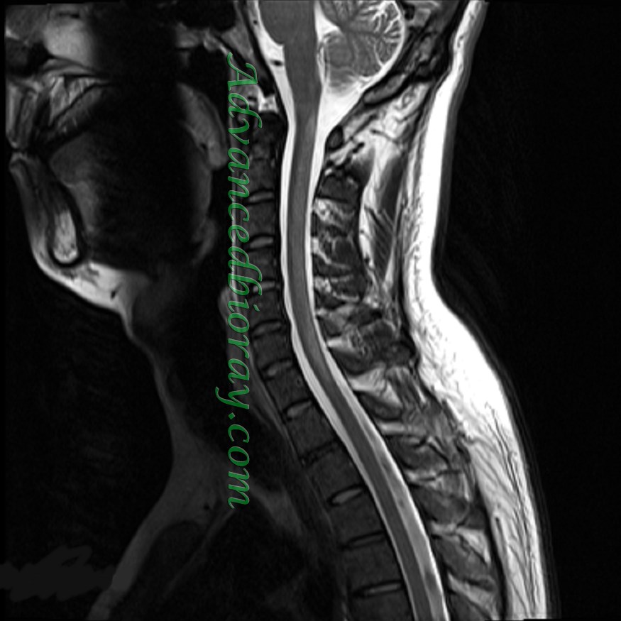
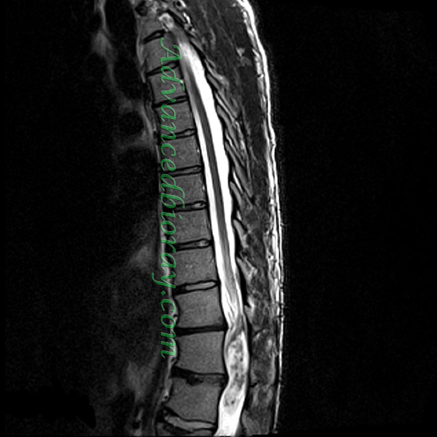
Case No 3 :
35-year-old man with clinical symptoms of anemia, paralysis, and pain.
MRI findings: Extensive signal abnormalities in the dorsal cord with a continuous pattern, without enhancement, consistent with B12 deficiency and associated transverse myelitis.
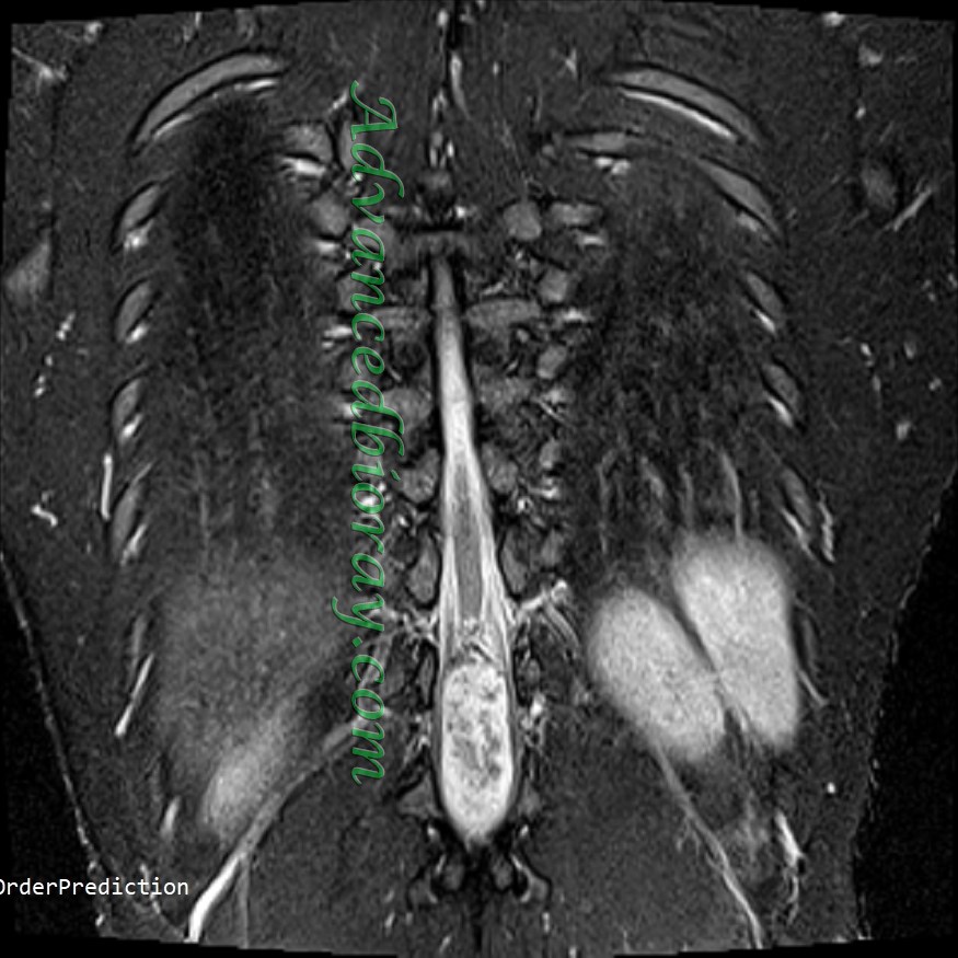
Case No 4 :
43-year-old man with progressive paralysis.
MRI findings: Abnormal heterogeneous hemorrhagic mass in the conus medullaris, consistent with ependymoma.