Magnetic resonance imaging (MRI) has revolutionized the diagnosis and evaluation of cerebrovascular disorders. It enables the early detection of ischemic infarctions, precise delineation of lesions near the skull base, and the identification of microangiopathy. MRI is particularly effective in diagnosing arteriovenous malformations and venous thrombosis without the need for contrast media. While computed tomography (CT) remains superior for the detection of acute hemorrhage, MRI excels in revealing intra-parenchymal and subarachnoid hemorrhages.
In recent years, the application of advanced dedicated sequences such as susceptibility-weighted imaging (SWI) has further enhanced MRI’s ability to provide detailed insights into vascular disorders. Additionally, advanced MRI techniques like magnetic resonance angiography (MRA) and spectroscopy hold the potential to replace conventional angiography and positron emission tomography (PET) for specific applications.
From our MRI archive , the following cases in the field of vascular disorders are shared:
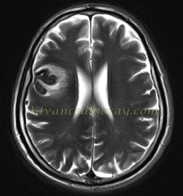
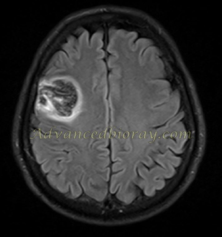
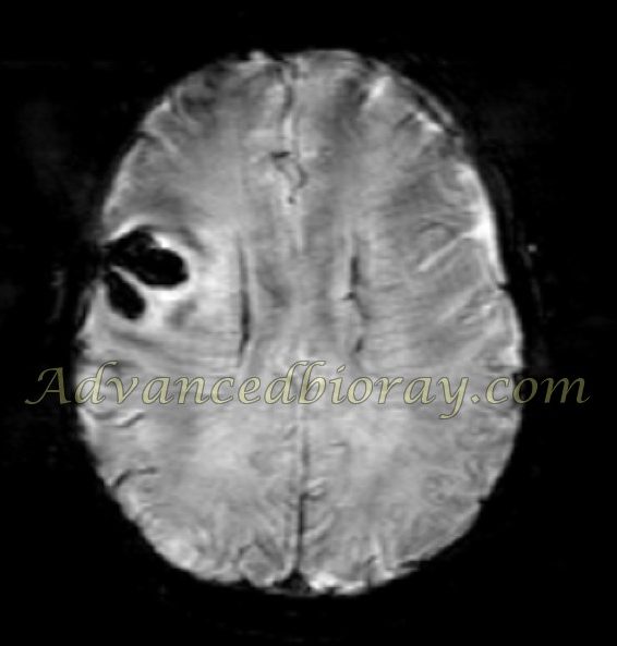
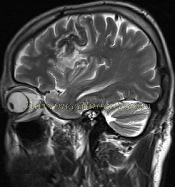
Case No 1
A 50-year-old woman presented with paresthesia. MRI revealed a large cavernoma on the left side of the pons. A signal void rim with hemosiderin deposition and a hemorrhagic center were observed.
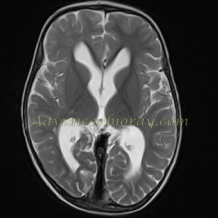
Case No 2
A 4-year-old boy with a history of sinus venous thrombosis underwent MRI with and without contrast. The imaging revealed a large filling defect in the superior sagittal sinus and the sinus confluence, which remained unenhanced on post-contrast sequences, consistent with the “empty delta sign.” Additionally, post-ischemic infarctions were observed in both periventricular regions, presenting as hyperintense signals on T2-weighted sequences.
Case No 3
A 65-year-old man presented with acute hemiplegia and aphasia. MRI demonstrated an acute gyral signal void abnormality in the cortex of the right parietal lobe, accompanied by superficial siderosis. The signal void appearance was prominently observed on susceptibility-weighted imaging (SWI) sequences.
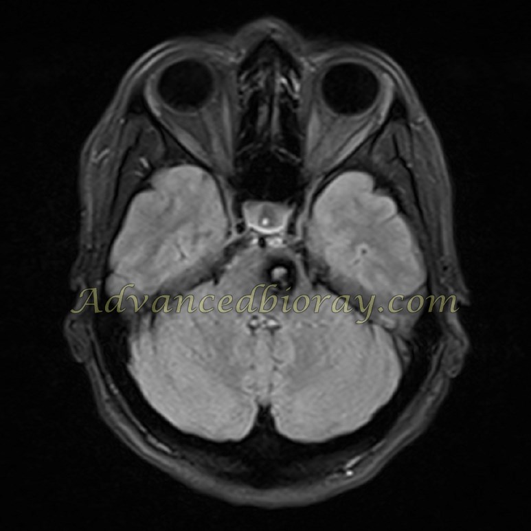
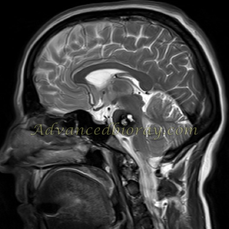
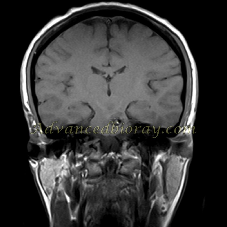
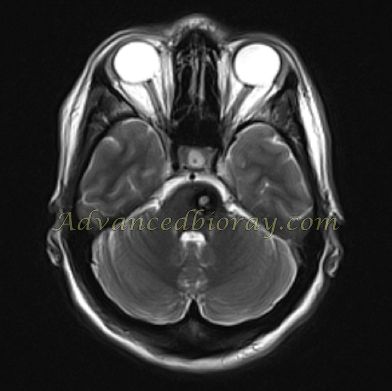
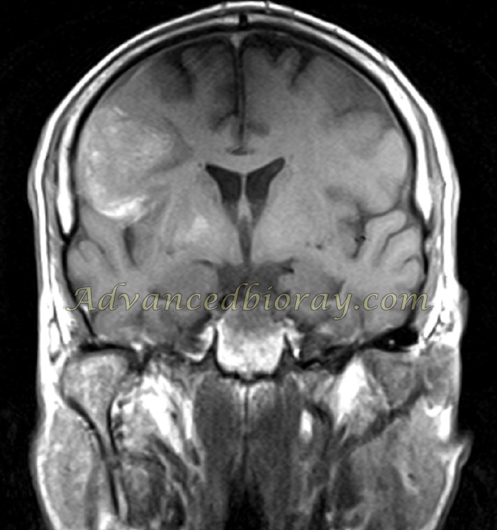
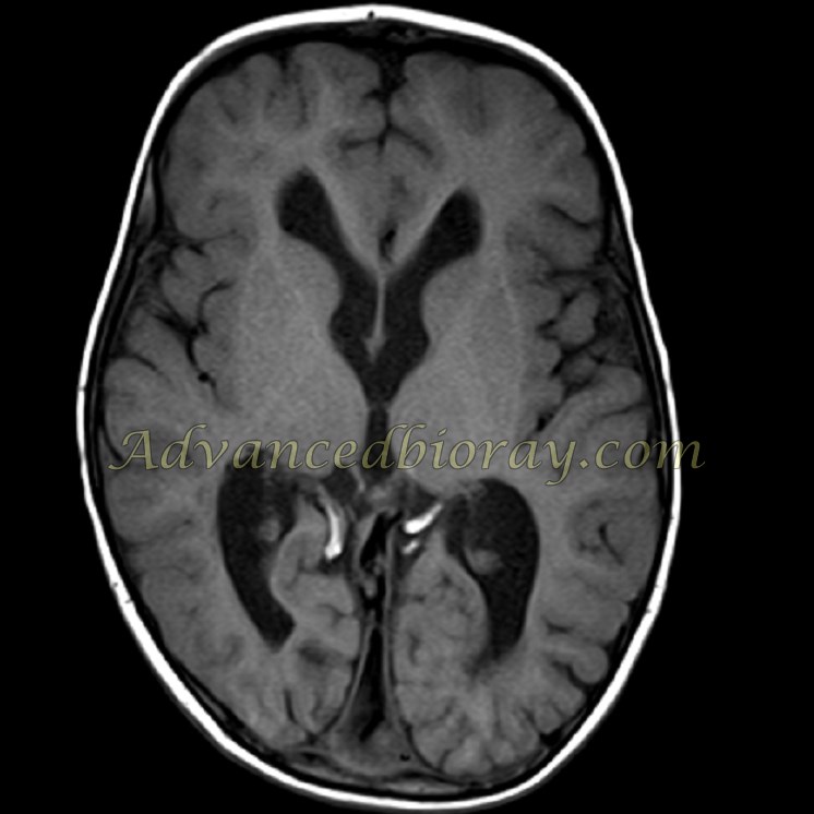
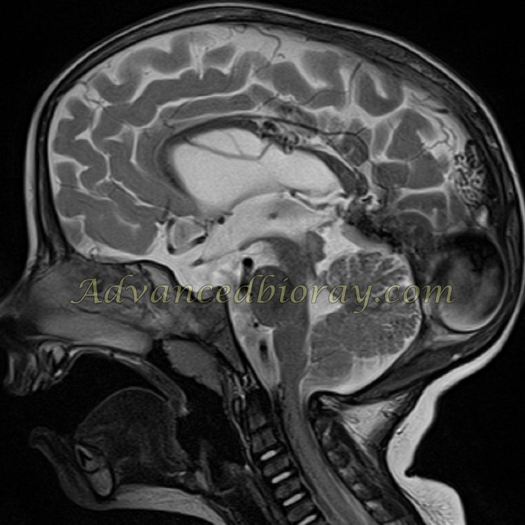
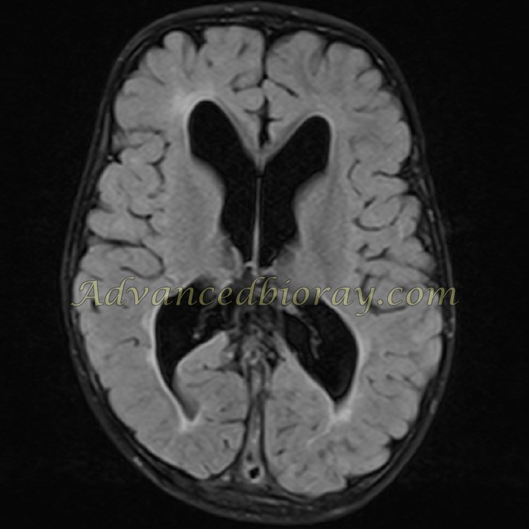
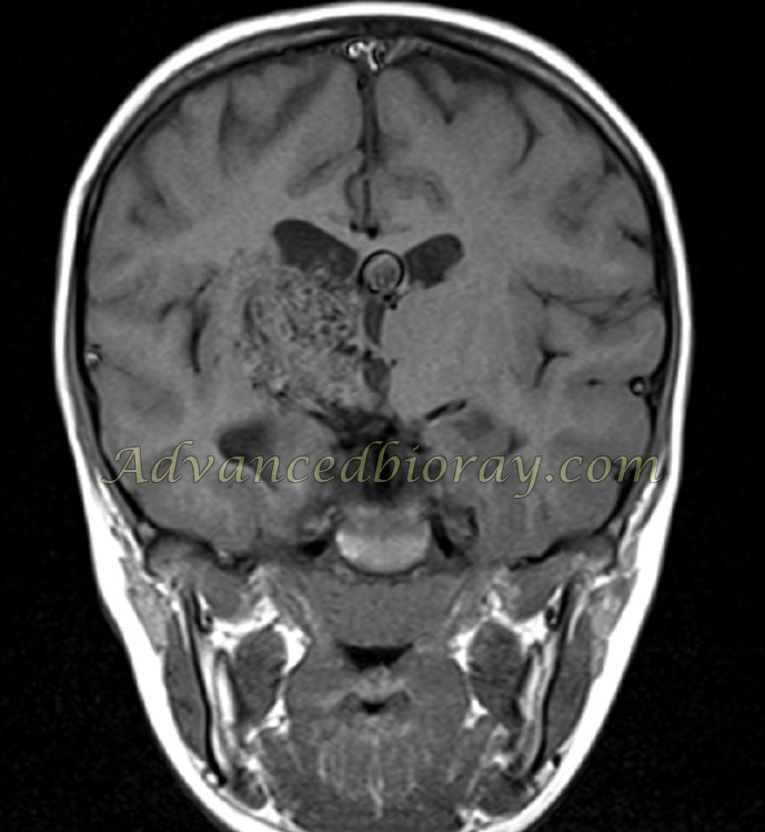
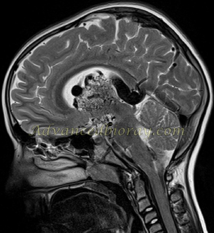
Case No 4
A 2-year-old boy with a seizure exhibits the typical appearance of an aneurysm of the vein of Galen (VGAM). The VGAM shows aneurysmal dilatation of the vein of Galen, with abnormal arterial feeders from the choroidal and thalamoperforating arteries, forming clusters of tortuous, dilated vessels. These vessels converge directly towards the vein of Galen without an intervening nidus, indicative of an arteriovenous fistula. The drainage of the vein of Galen is via a prominent straight sinus to the torcula of Herophili and the superior sagittal sinus.
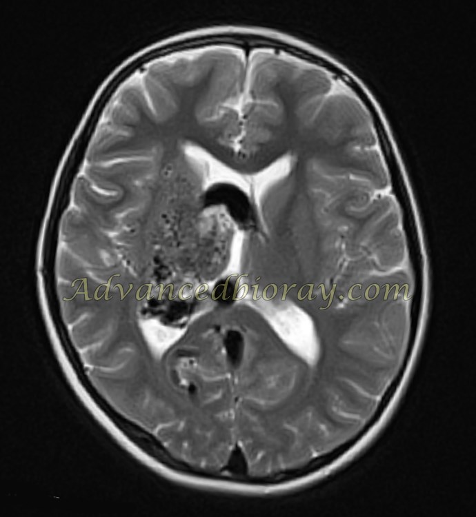
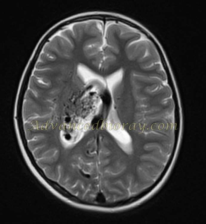
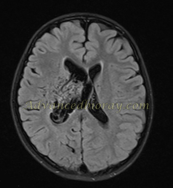
Case No 5
A 9-year-old girl with a seizure presents with an MRI showing a large arteriovenous malformation (AVM) in the right periventricular region. The AVM is associated with prominent feeding arteries and a cluster of abnormal small vessels. Surrounding brain tissue appears gliotic.