Epilepsy is one of the most common serious brain conditions, affecting over 70 million people worldwide. Its incidence has a bimodal distribution with the highest risk in infants and older age groups. With expansion of neuroimaging and its ability in diagnosis of small FCD and other lesions in Brain, new windows are opened for patients’ relief from this long-lasting process.
Advances in brain imaging are helping to identify the structural and functional causes and consequences of the epilepsies. In neuroimaging not only the anatomic location of lesion is identified but also fMRI, DTI, MRS and VBM as well as new post processing options before and after operation play great role is diagnosis and treatment.
More than 25000 cases have been gathered since 2003 till now and categorized ready for scientific collaborations. Also, a group of Well-known Epileptologists are in the scientific committee and ready for second opinion for the patients who doesn’t have access to medical professional team in Epilepsy.
Some of the cases are introduced here :
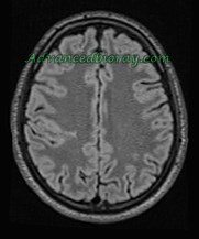
Case No 1
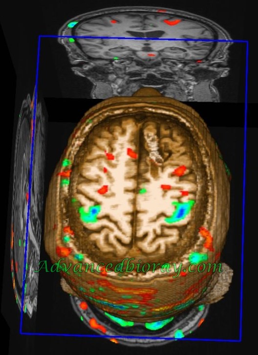
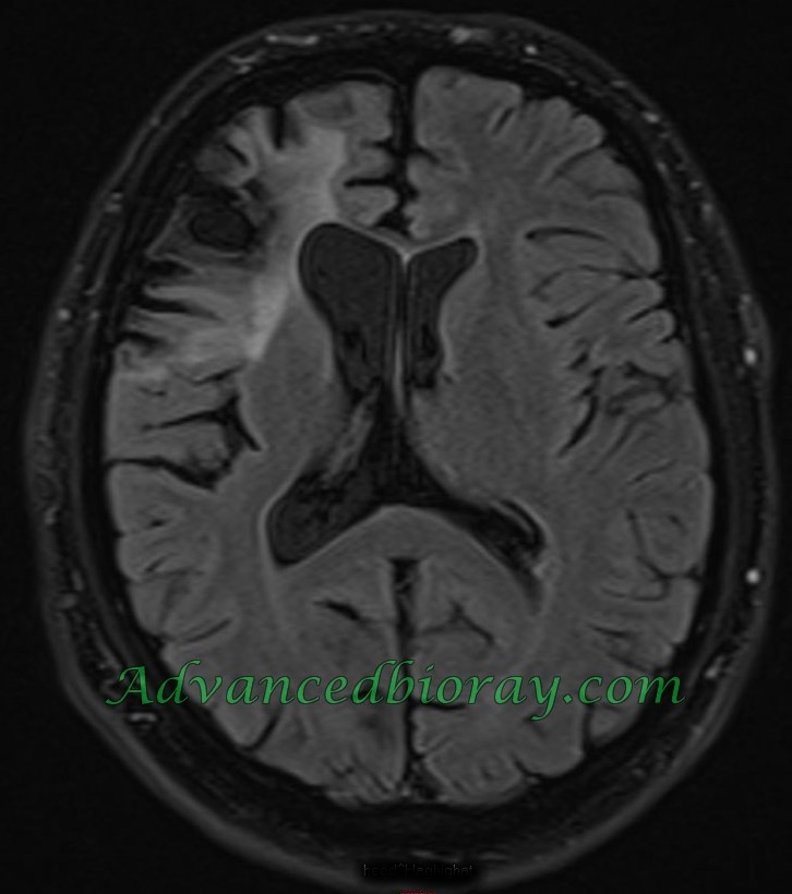
17-year-old boy with history of focal epilepsy in the MRI and especially in the VBM there is typical FCD Type IIB in the right fronto parietal cortex with typical Radiation band. The extension of FCD Type IIB is well detectable by morphometric study.
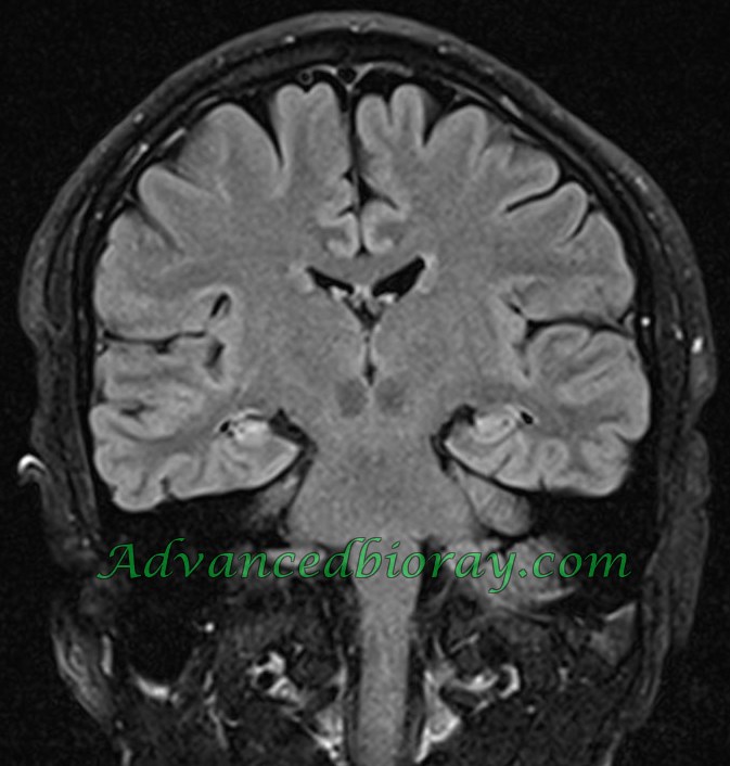
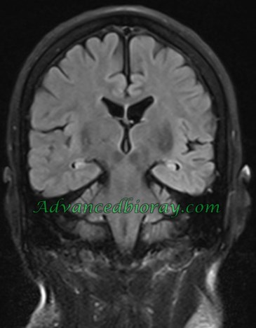
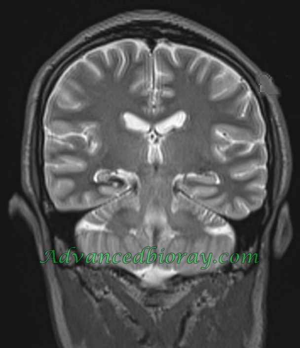
Case No 2
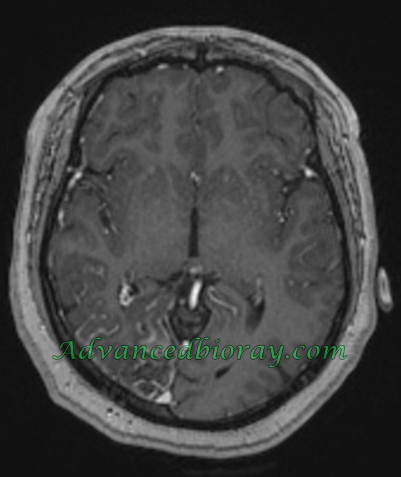
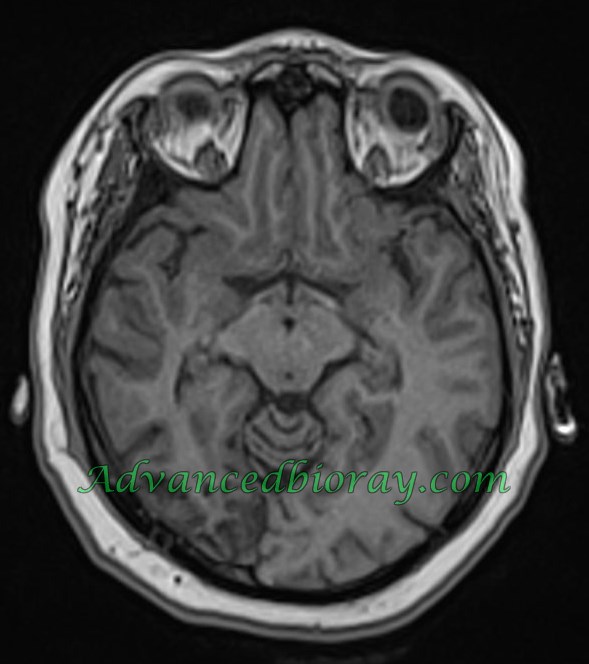
30 year old man with focal temporal lobe epilepsy representing right Amygdalla and hippocampal sclerosis .
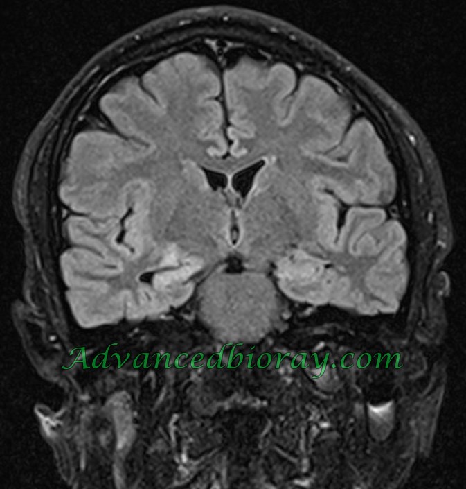
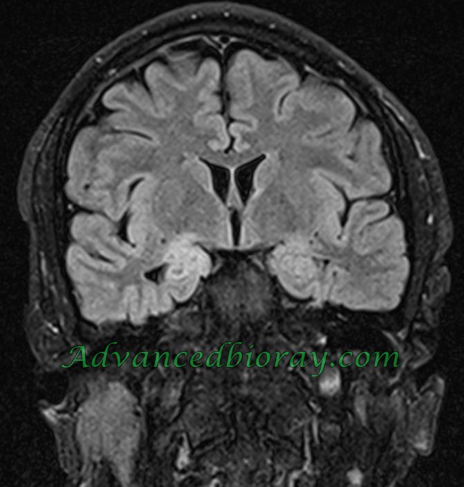
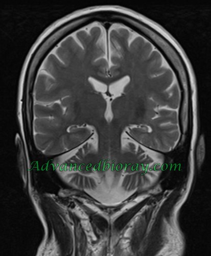
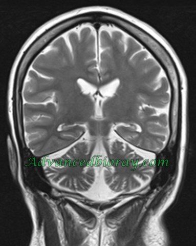
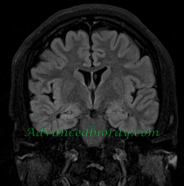
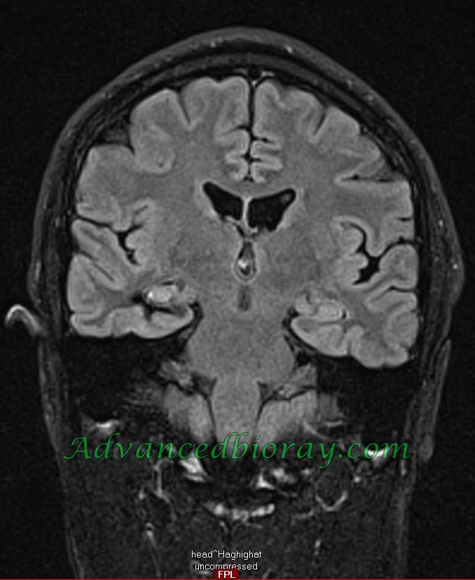
Case No 3
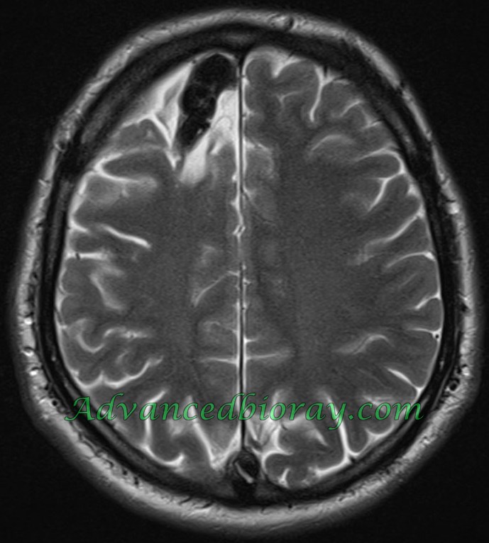
A 53 year old right-handed female presented with a history of intractable seizures since her childhood . In the MRI bilateral hippocampal sclerosis and atrophy of cerebellum is seen .
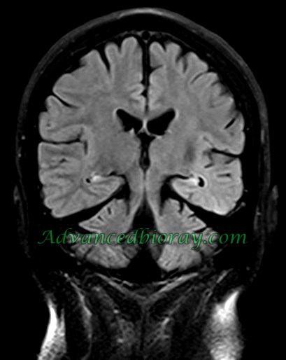
Case No 4
A 29-year old right-handed female presented with history of seizure at the age of 2. MRI represented dual lesion as right sided hippocampal sclerosis and “Sturge -Weber syndrome ” with right sided pial leptomeningial angiomatosis .
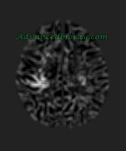
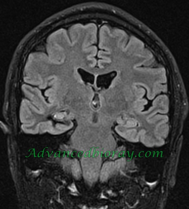
Case No 5
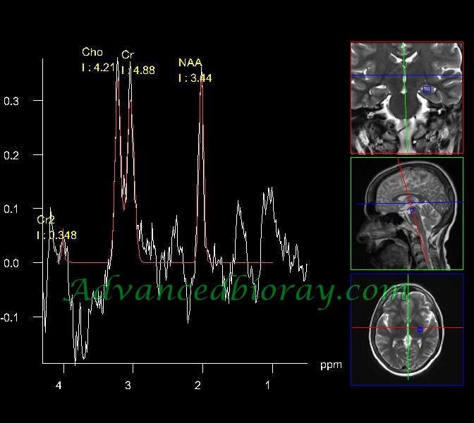
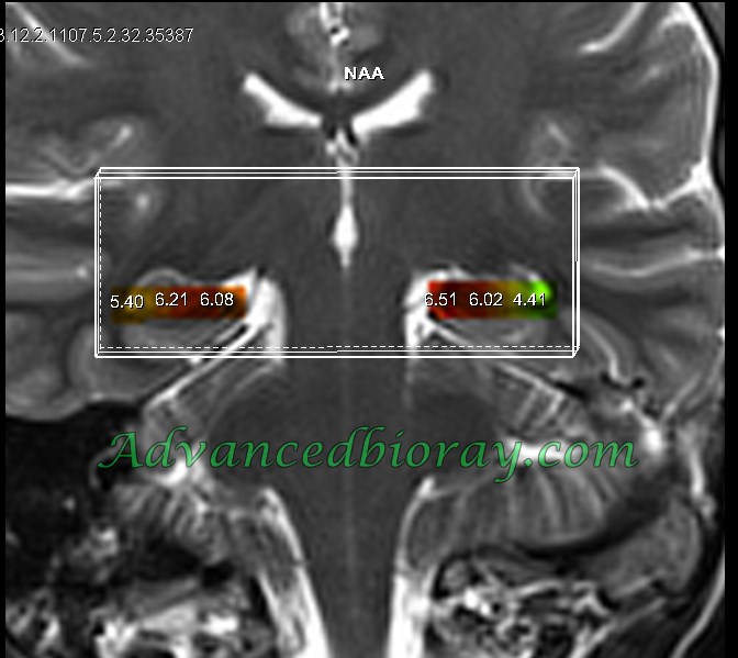
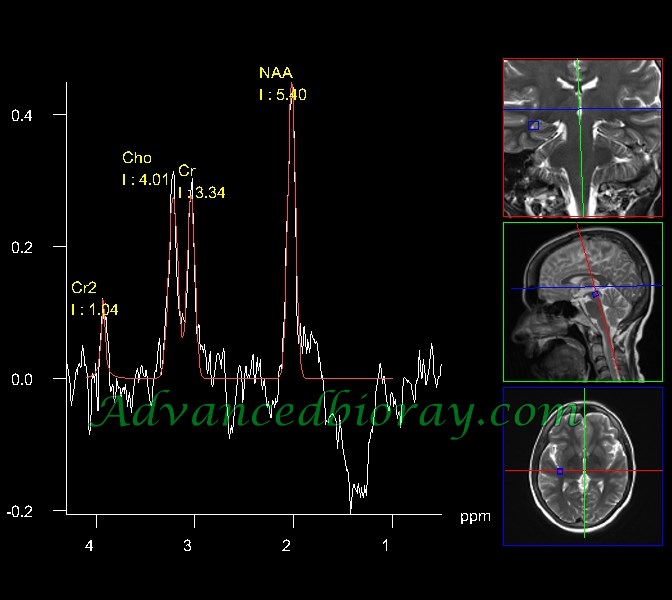
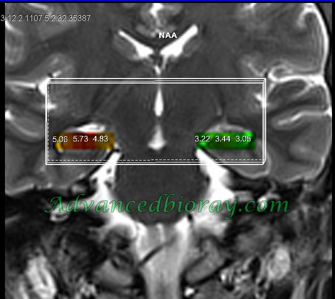
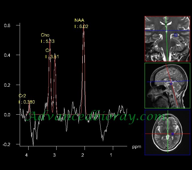
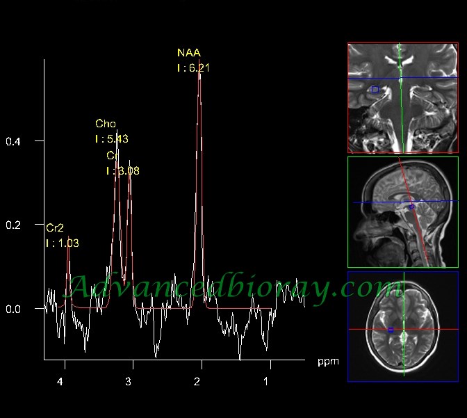
20 year old woman with history of left temporal lobe refractory seizure ,In the MRI left sided hippocampal sclerosis is seen and in the correlated MRS ,there is reduction in the NAA/Cr level confirming neuronal loss and gliosis .
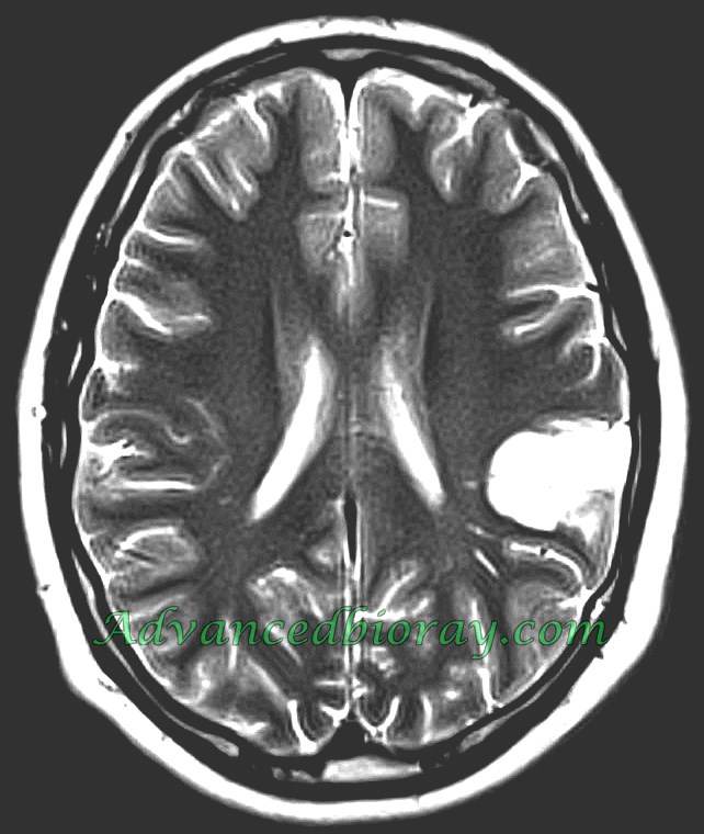
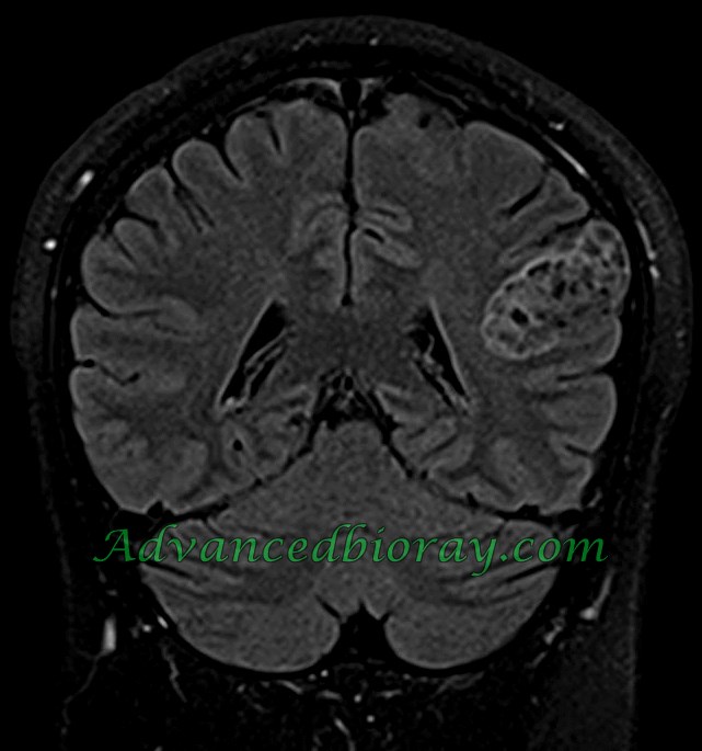
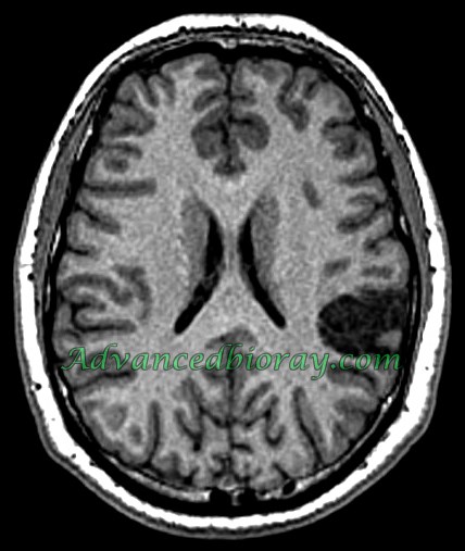
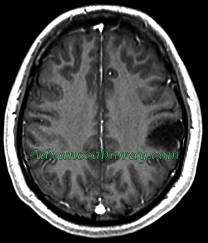
Case No 6
10-year-old boy with refractory seizure representing a cortical located tumoral mass in the left tempor- parietal cortex which is unenhanced and has typical “Blubby appearance “.
Dysembryoplastic neuroepithelial tumors (DNET) is confirmed after operation.
Case No 7
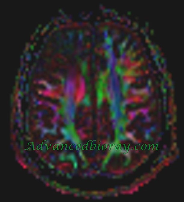
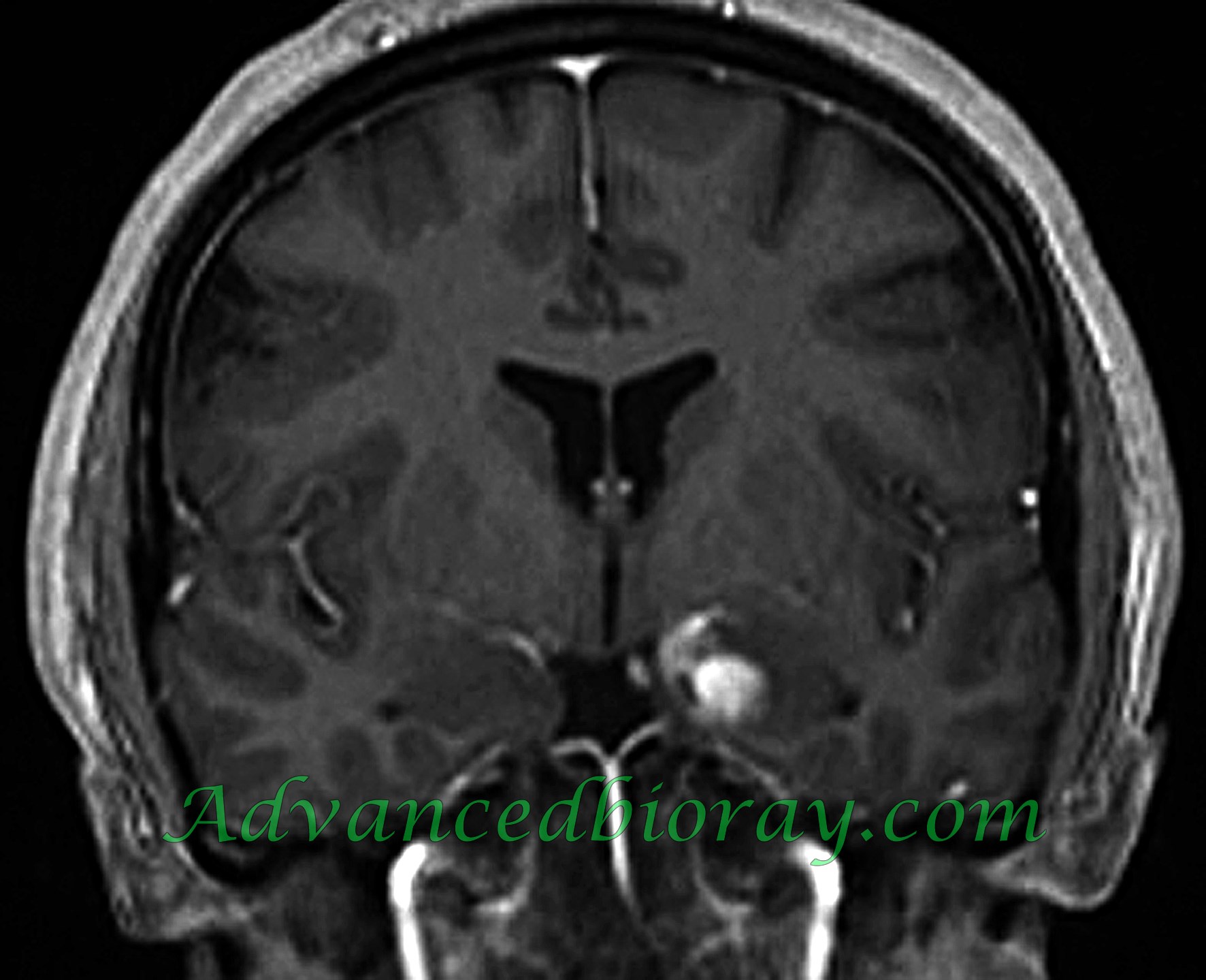
a 42-year-old right-handed male presented with history of seizure from 26 years ago after penetrating war injury (in 1982 with 10-day-LOC). Attacks started with experiential aura (anxiety), left head and eyes deviation then secondary generalization followed by postictal confusion and left side hemi paresis. Tracing showed many right-side epileptiform discharges mainly on right frontocentral and temporal region. MRI represented extensive brain malasia and signal void abnormal Tumifactive lesion with destruction of normal fibers . In post surgical evaluation and based on pathology Foreign body with sever malasia was confirmed .
Case No 8
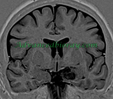
16 year old girl with left temporal lobe refractory seizure ,MRI represents abnormally enhanced mass in the left deep temporal lobe with typical configuration in favor of Ganglioglioma.
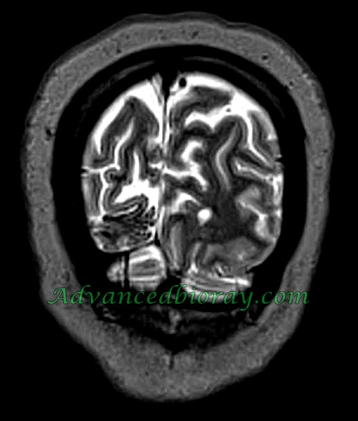
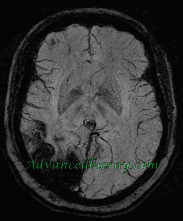
Case No 9
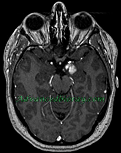
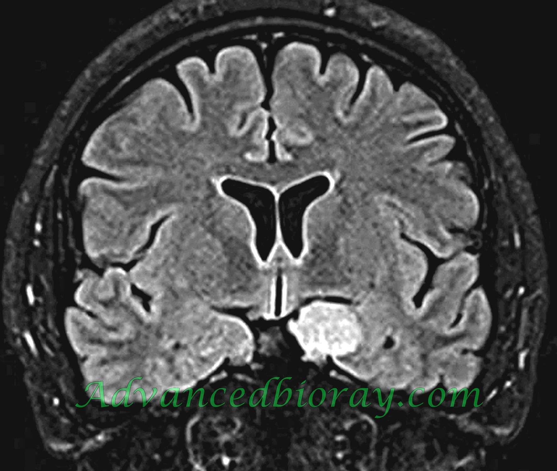
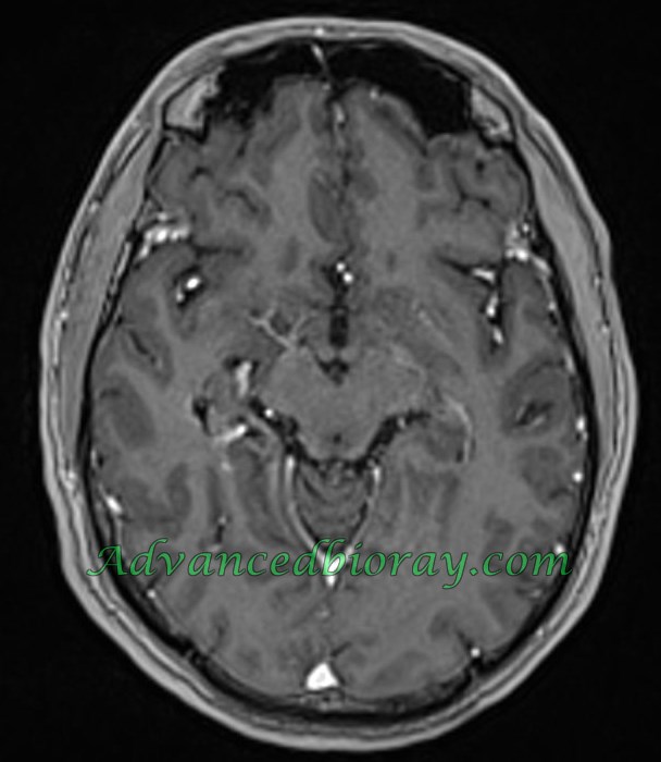
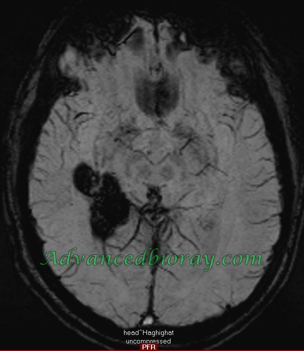
32 year old man with focal Epilepsy and represented Cavernoma in the right temporal lobe . In the correlated SWI sequence extensive hemosiderin deposition is detectable . There is associated right sided hippocampal sclerosis .
Case No 10
39-year-old woman with history of focal epilepsy and normal conventional MRI, but correlated morphometric studies with post processing and VBM represents high signal area in the right frontal lobe operculum and confirmed after operation as FCD Type I .