Musculoskeletal system imaging is one of the important Fields in the medical diagnostic imaging.
It has crucial role in diagnosis and follow up of patients with Bone, Muscles, Ligaments and tendons as well as Arthropathies and soft tissue disorders. Different imaging modalities are used in the field including Simple X-Ray, CT stan, Ultrasonography and MRI. In the field of trauma and Emergency mostly simple Xray and CT scan are modality of choice however, for detail discrimination of soft tissue surrounding joints MRI is modality of choice. Nowadays before every operation of ligaments and Bony of soft tissue lesions MRI has very important role. During over 15-year daily MRI with many cases of MSK with different diagnosis are gathered as a rich archive suitable for teaching file and researches. Here we introduce some of our cases:
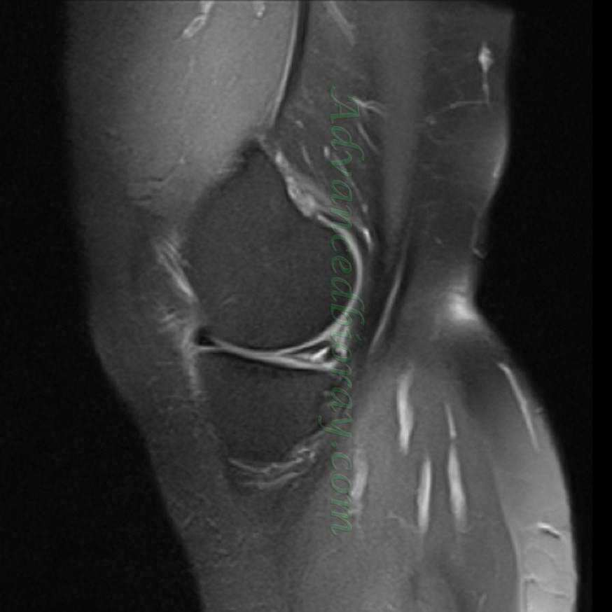
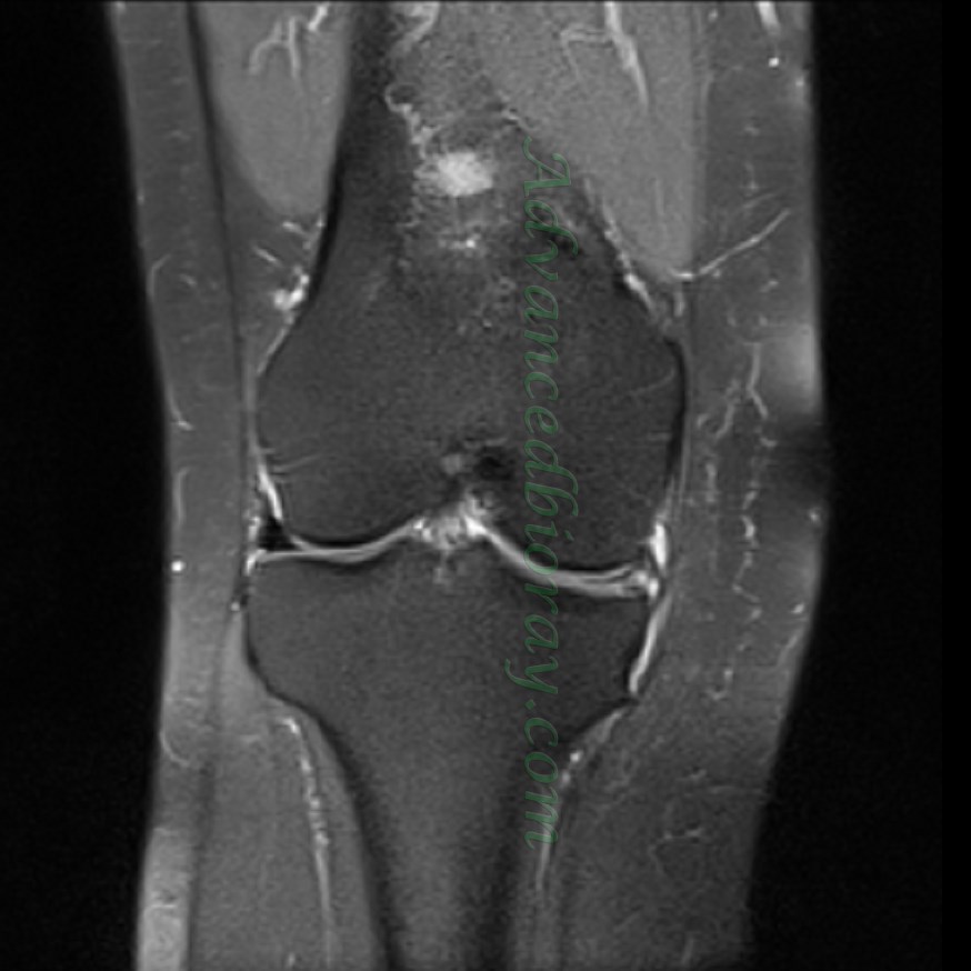
Case No 1
The patient is 54-year-old woman with a history of locking the knee, In the MRI there is a horizontal tear in the body and posterior horn of the medial meniscus with cystic formation. This important finding is confirmed by coronal, axial, and Sagittal PD-FS images.
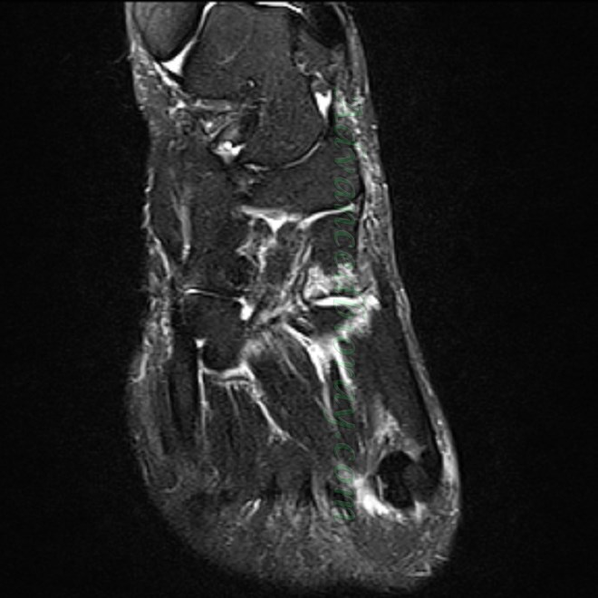
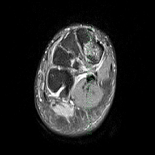
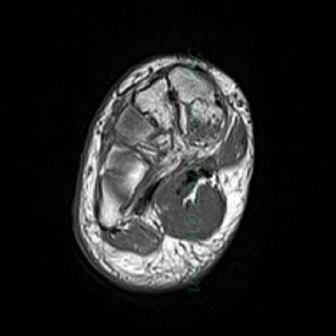
Case No 2
The patient is a 40-year-old man referred with ankle pain; In the obtained MRI, Stress fracture in the medical cuneiform bone is detectable in all sequences including Axial T1W and Axial, Coronal images in PD-FS sequence.