Neurodegenerative Disorders: Overview and Case Studies
Neurodegenerative disorders, including Alzheimer’s disease (AD) and Parkinson’s disease (PD), are among the most common and devastating conditions affecting patients and their families. Although symptomatic therapies are available, they are often inadequate. For example, therapies for PD provide only partial relief, while those for dementia offer modest benefits at best. Moreover, current treatments do not significantly alter the relentless progression of these disorders.
Advances in basic neuroscience have raised hopes for disease-modifying therapies, which are likely to be most effective when applied early in the disease course. Early detection would enable the administration of these therapies to those who would benefit most, at the stage when they are most likely to succeed. Additionally, early diagnosis facilitates the selection of appropriate participants for clinical trials of novel treatments and excludes those unlikely to benefit. It also allows for better prognostic predictions and the optimal use of healthcare resources.
Neuroimaging techniques hold significant promise for the early diagnosis of neurodegenerative disorders. These techniques can either distinguish specific diseases from other conditions with similar early-stage presentations or detect central nervous system dysfunction before clinical symptoms emerge. The latter approach is particularly useful in populations at increased risk of developing neurodegenerative diseases, helping identify individuals for neuroprotective trials or, eventually, for disease-modifying therapies.
In our image database, numerous cases of neurodegenerative disorders have been archived for research purposes. Below are examples of diagnosed cases from our archive:
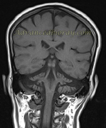
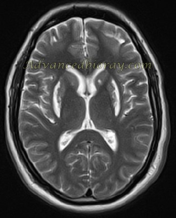
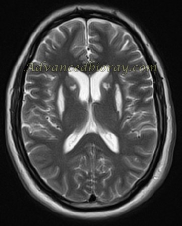
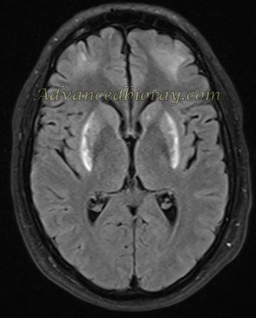
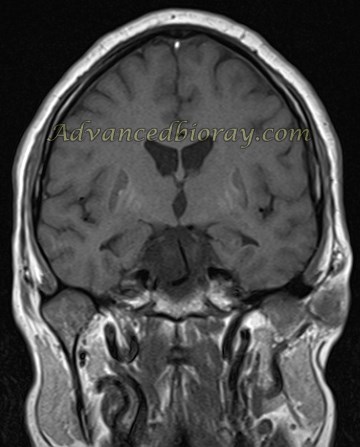
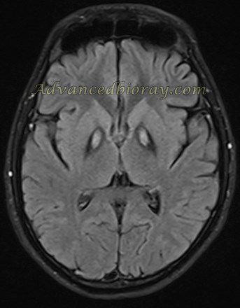
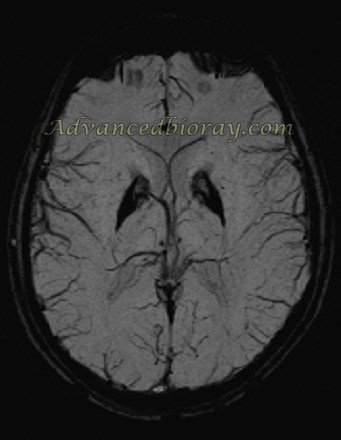
Case No. 1
Axial T2-weighted and coronal T1-weighted MRI images of a known case of dementia show an MTA score of grade 3 on the left side, along with asymmetrical left temporal lobe atrophy. These findings are consistent with dementia.
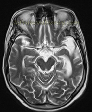
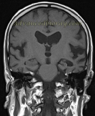
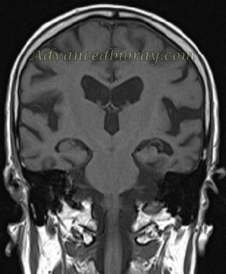
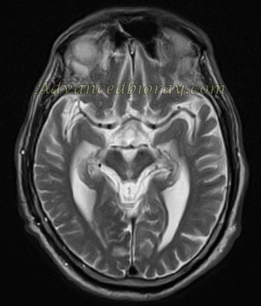
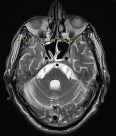
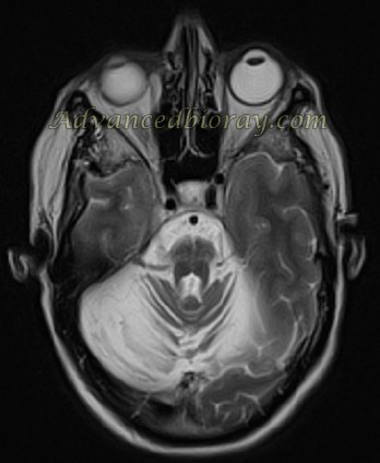
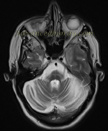
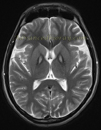
Case No. 2
A known case of Parkinson’s syndrome with MRI findings of significant cerebellar atrophy, the characteristic “hot cross bun” sign, and pontine atrophy. The bilateral middle cerebellar peduncle (MCP) diameter measures 8 mm, suggestive of MSA-C (multiple system atrophy, cerebellar type).
Case No. 3
A 32-year-old man with a history of methanol intoxication. Axial T2-weighted and FLAIR images show bilateral symmetrical putamen necrosis 15 weeks after intoxication. Coronal T1-weighted images reveal abnormal high signal intensity in the basal ganglia, consistent with necrosis and hemorrhage.
Case No. 4
Axial T2-weighted, T1-weighted, and SWI images of a case diagnosed with an iron deposition disorder. The images demonstrate the typical “tiger eye” appearance, characterized by signal voids and iron deposition in the globus pallidus.
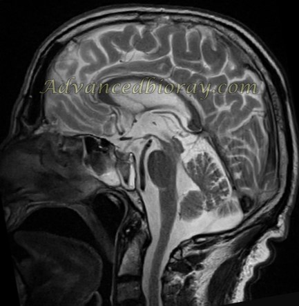
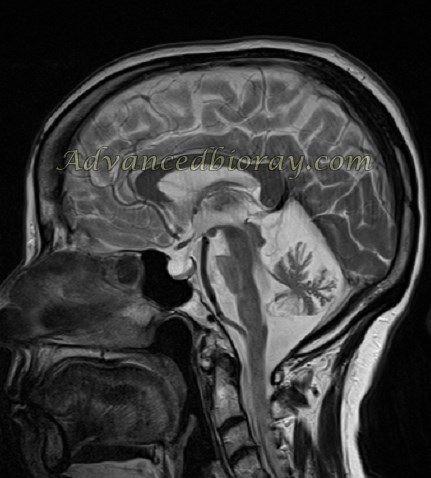
Case No. 5
A 46-year-old man with parkinsonism. MRI evaluation of the brainstem and posterior fossa metrics, supported by the Parkinson index, reveals significant atrophy in the bilateral superior cerebellar peduncles and a reduced anteroposterior diameter of the brainstem.The characteristic “hummingbird sign” is present, strongly suggesting PSP (progressive supranuclear palsy).