Magnetic resonance imaging (MRI) plays a vital role in diagnosing and managing pediatric neurometabolic disorders. With its high sensitivity and specificity, brain MRI can predict inherited neurometabolic conditions by identifying key findings such as bilateral symmetric white matter abnormalities and diffusion restriction in deep grey nuclei.
MRI provides essential insights for classifying these disorders into dysmyelinating and demyelinating categories, while magnetic resonance spectroscopy (MRS) complements the process with additional metabolic data critical for diagnosis and follow-up. Early and accurate diagnosis is crucial, as some disorders have treatment options that can prevent severe neurological consequences or even mortality.
Advanced MRI techniques, including diffusion-weighted imaging and MRS, enable precise differentiation of neurological conditions such as ischemic, hypoxic, inflammatory, and metabolic diseases in infants and children. Neuroradiologists must be well-versed in identifying these disorders to ensure timely intervention and avoid the risks associated with delayed diagnosis.
With over 15 years of experience specializing in pediatric MRI/MRS, our group has developed an extensive and invaluable archive in this field. Below are some notable cases:
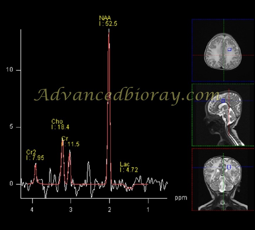
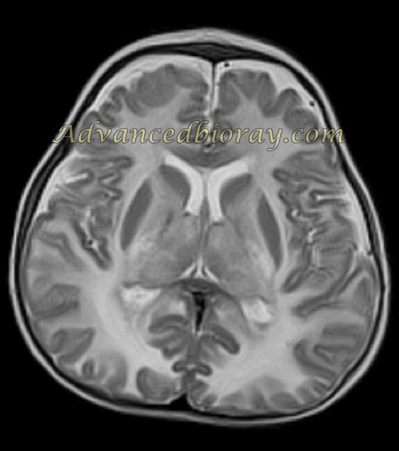
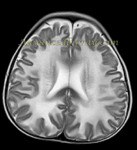
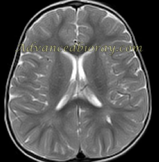
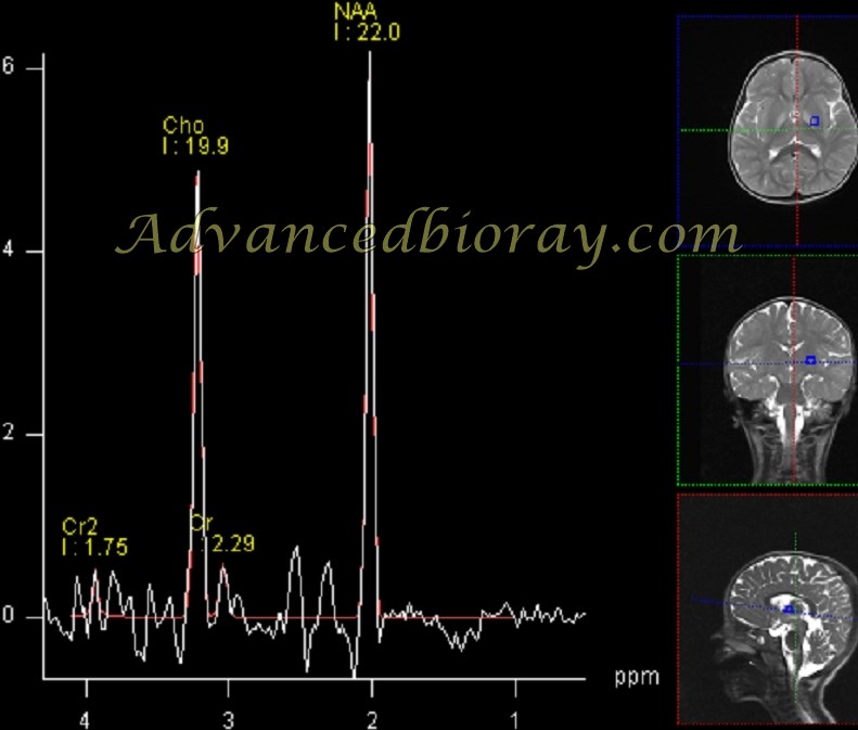
Case 1: Canavan Disease
A 6-month-old boy presented with macrocephaly, seizures, and dystonia. MRI revealed extensive white matter signal abnormalities with typical involvement of the globus pallidus and no involvement of the caudate and putamen. Magnetic resonance spectroscopy (MRS) demonstrated a prominent NAA peak, characteristic of Canavan disease.
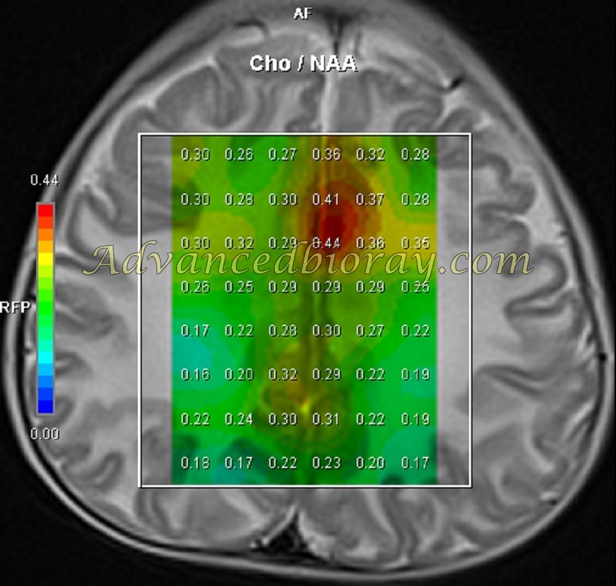
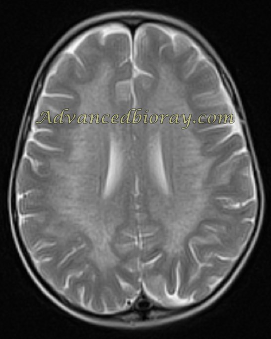
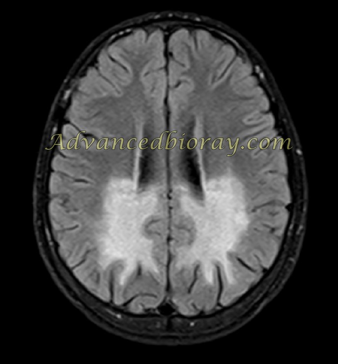
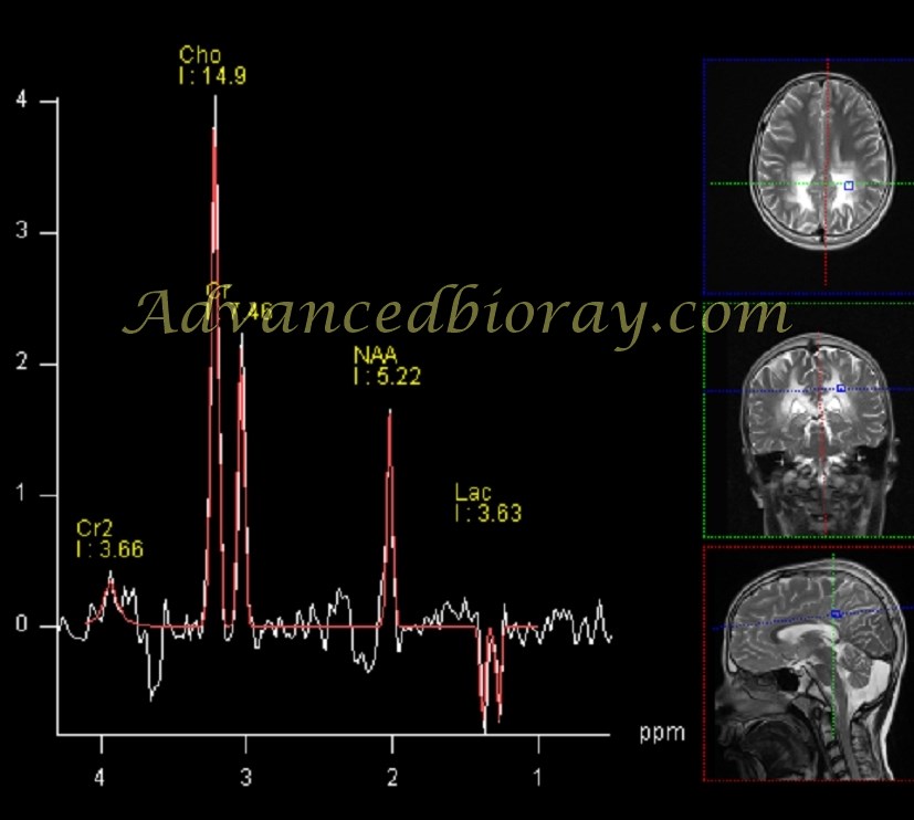
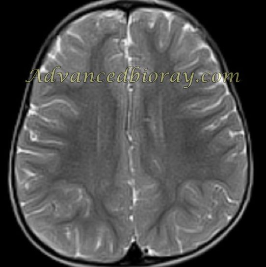
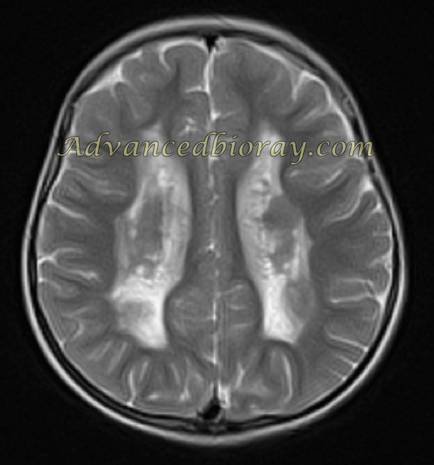
Case 2: Metachromatic Leukodystrophy (MLD)
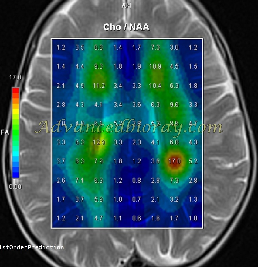
A known case of metachromatic leukodystrophy (MLD) presented with typical white matter signal abnormalities in both centrum semiovale. Correlated MRS findings showed an elevated Cho/Cr ratio, indicative of the demyelination process.
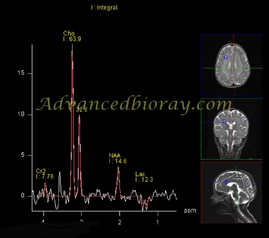
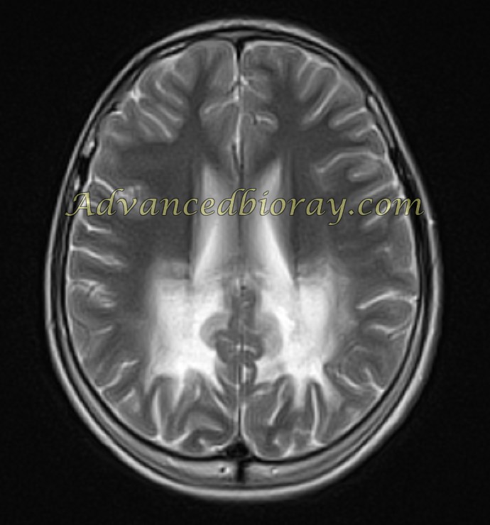
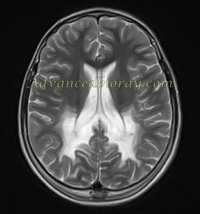
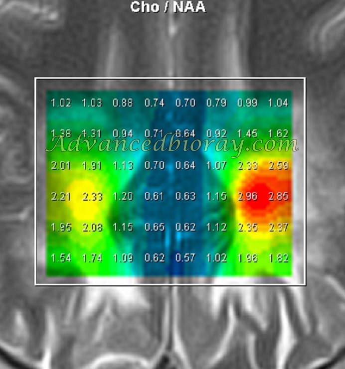
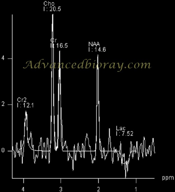
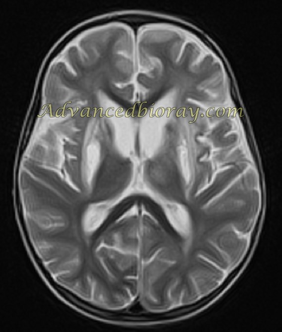
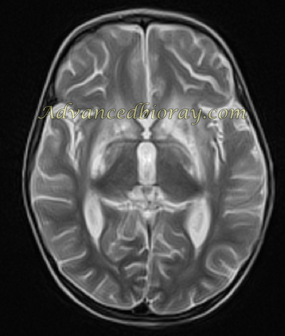
Case 3: Adrenoleukodystrophy (ALD)
A known case of adrenoleukodystrophy (ALD) showed more pronounced involvement of the bilateral parietal white matter on MRI. MRS findings revealed a typical rise in choline and lactate levels, consistent with the demyelination process.
Case 4: Creatine Deficiency
A known case of creatine deficiency presented with a normal MRI appearance. However, MRS findings in long TE sequences revealed the absence of the normal creatine (Cr) peak, confirming the diagnosis.
Case 5: Leigh Disease
A known case of Leigh disease exhibited symmetric T2-weighted hyperintense lesions in the basal ganglia (putamen, globus pallidus, and caudate), thalami, midbrain (red nucleus, substantia nigra, and periaqueductal region), brainstem, and dentate nuclei. MRS showed a typical rise in lactate, confirming the diagnosis.
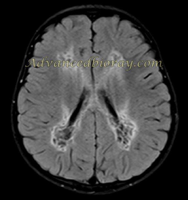
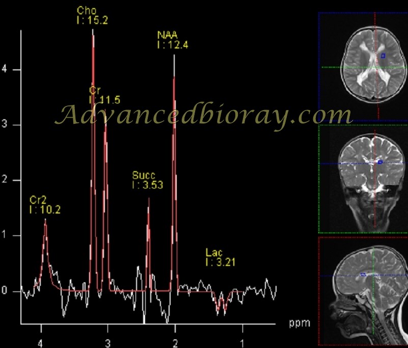
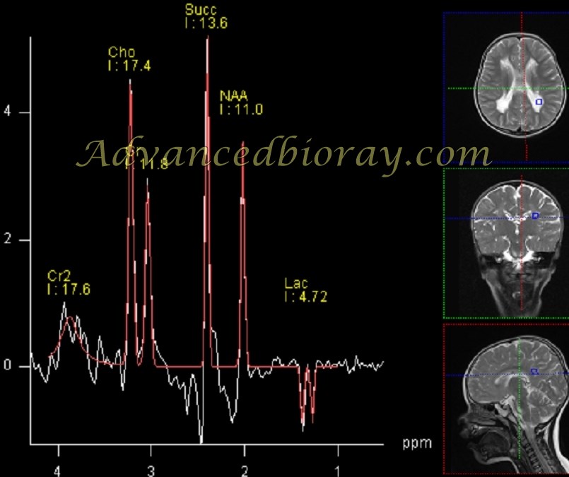
Case 6: Succinate Dehydrogenase Deficiency (SDHD)
Known cases of SDHD, a typical succinate peak in MRS, confirms the diagnosis.