Brain tumors are the most common form of solid tumors in children and the leading cause of death from solid tumors in this age group. Most brain tumors in children are primary CNS lesions. MR imaging is the modality for diagnosing and evaluating intracranial neoplasms in children. It is ideal for initial preoperative diagnosis, assessing tumor extent, treatment planning, and image-guided therapies. Advanced imaging techniques, such as MRS, PWI, fMRI, and DTI, play a significant role in diagnosis and follow-up. With over 20 years of experience in neuroimaging, we have a dedicated archive of pediatric brain tumors classified based on pathology. This provides a great research opportunity, both nationally and internationally. We welcome anyone interested in this field and introduce some of our cases below:
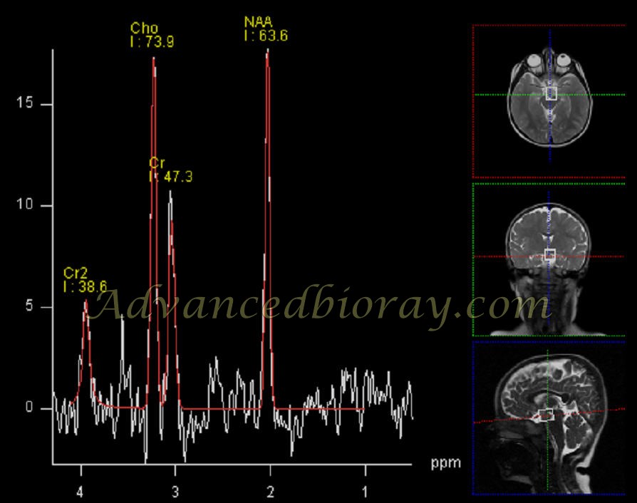
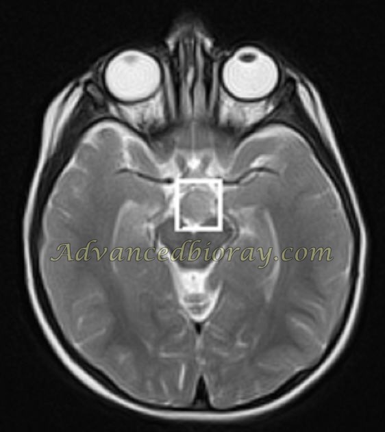
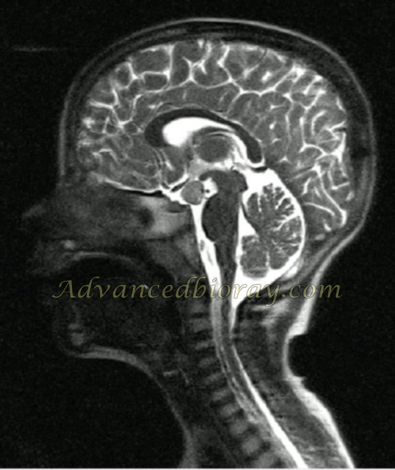
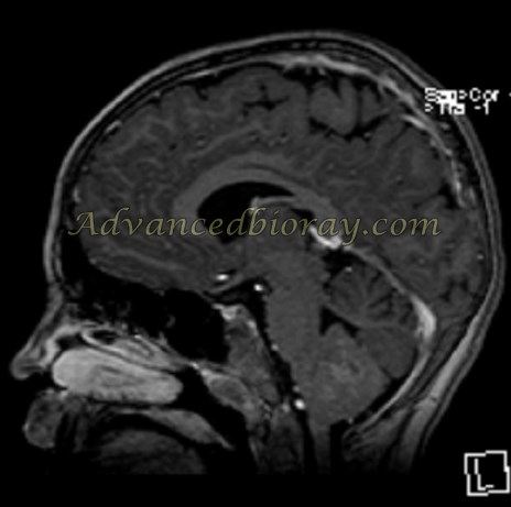
A 16-month-old girl with an abnormal mass in the tuber cinereum, typical of hypothalamic hamartoma, with MRS showing an increased Cho/NAA ratio.
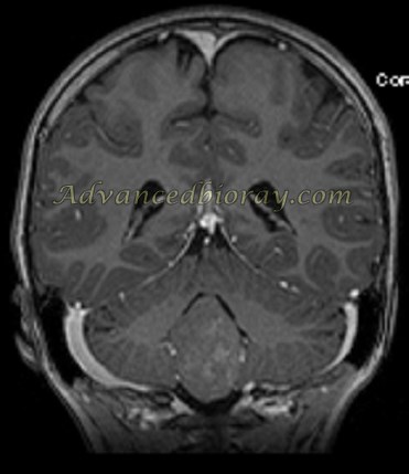
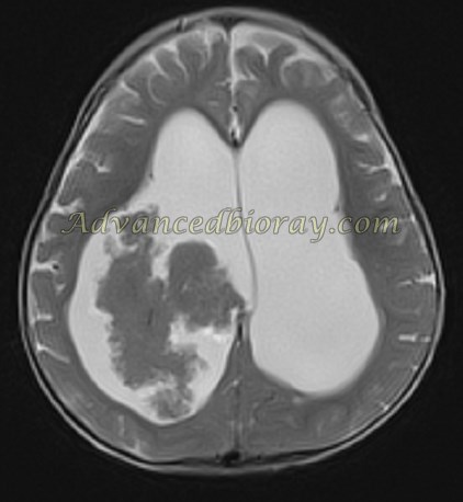
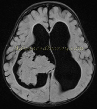
A 4-year-old boy with an enhancing mass in the floor of the fourth ventricle, diagnosed as Ependymoma.
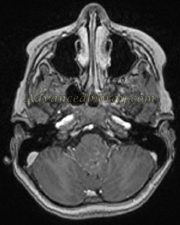
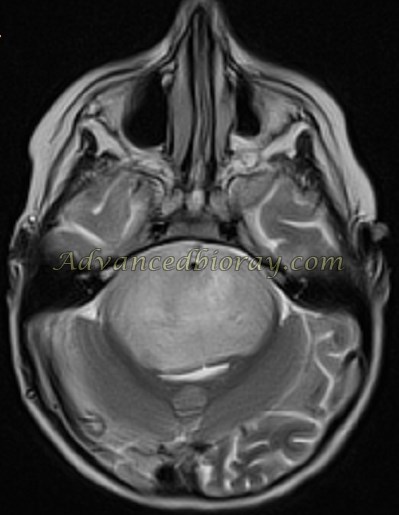
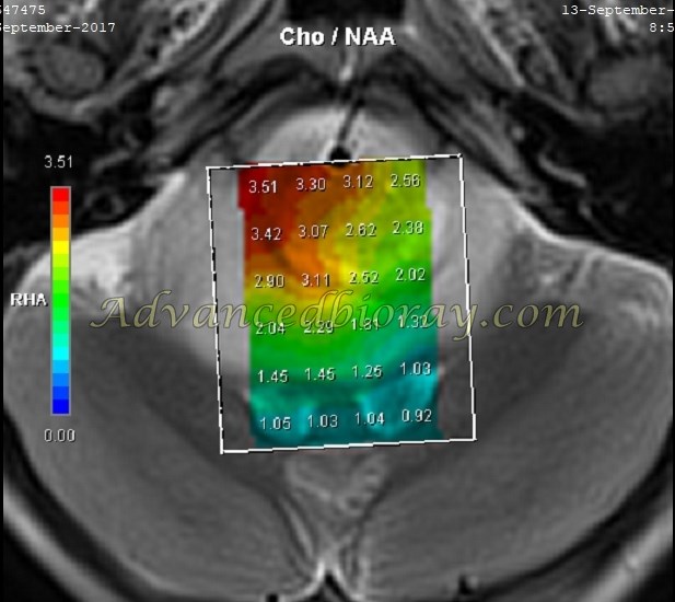
A typical Pontine Glioma with an MRS profile consistent with high-grade glioma.
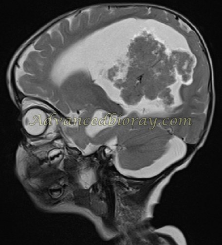
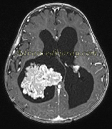
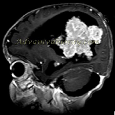
A 1-year-old boy with a large enhancing intraventricular Choroid Plexus mass.
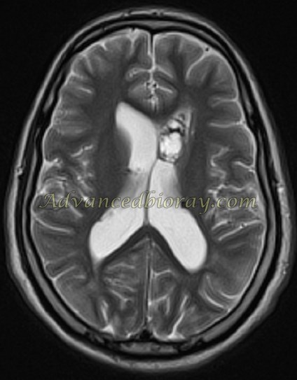
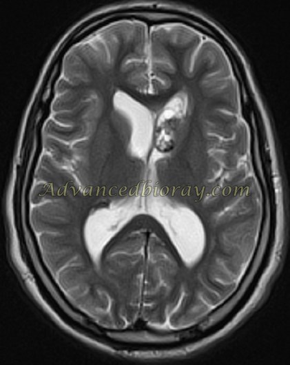
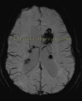
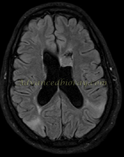
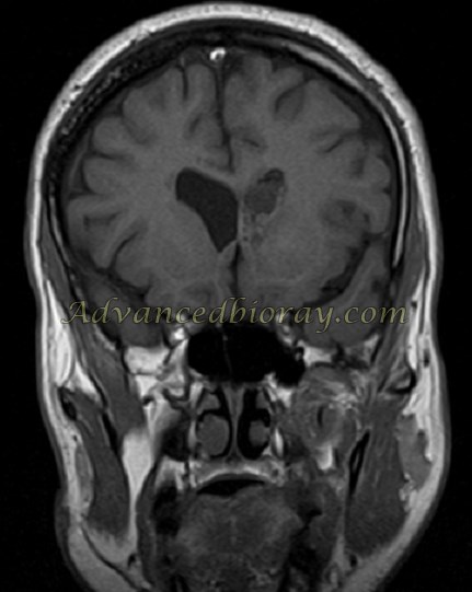
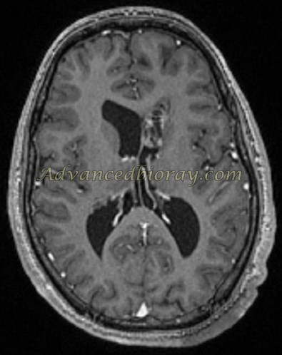
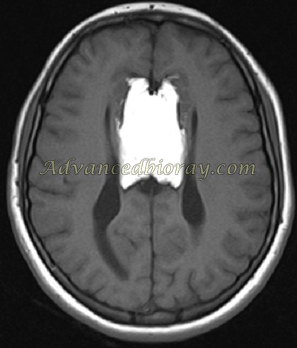
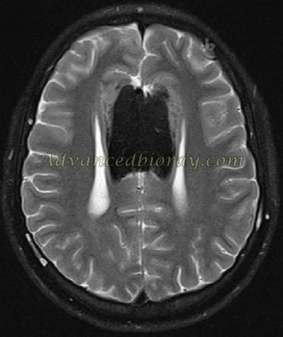
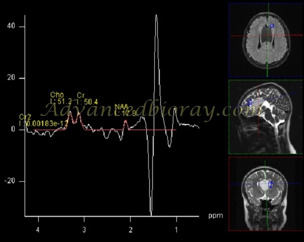
A 15-year-old boy with a full presentation of tuberous sclerosis (TS), showing subependymal calcified nodules and subcortical hamartomas, superimposed by an abnormally enhanced calcified mass near the left foramen of Monro, consistent with giant cell astrocytoma.
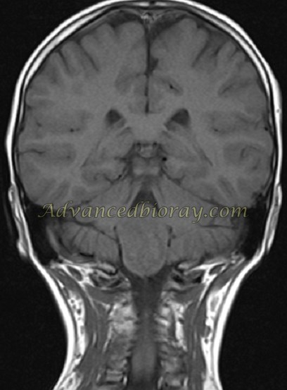
A spectrum of midline commissural anomalies with hypoplastic changes in the corpus callosum and midline lipoma, with specific MRS findings showing a lipid peak.