Epilepsy is one of the most common disorders of the nervous system, affecting children and adults across all races and ethnic backgrounds. A seizure occurs when one or more parts of the brain experience a surge of abnormal electrical activity, disrupting normal brain function. There are two primary categories of epileptic seizures: focal (partial) seizures and generalized seizures.
Imaging plays a crucial role in the diagnosis, treatment, and follow-up of patients with epilepsy. With over 20 years of experience and more than 20,000 cases in epilepsy diagnosis, our group is well-prepared to pursue further investigations in various areas related to epilepsy. The introduction of advanced software, artificial intelligence (AI), and post-processing techniques makes it an even more attractive field for researchers from different disciplines.
Also, we introduce the link for the book of neuroimaging in Epilepsy which was written 6 years ago and contains images from our cases who were proved with prognosis and pathology report. We introduce some of samples here:
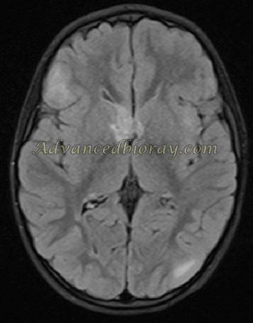
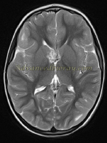
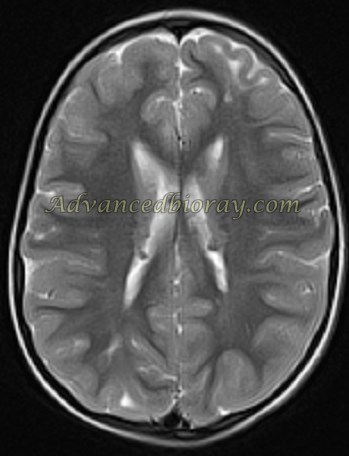
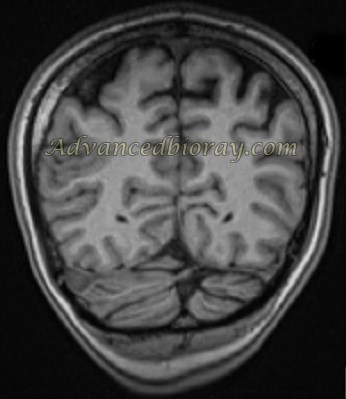
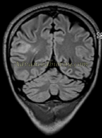
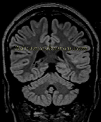
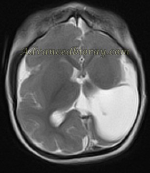
4-year-old girl with epilepsy full-presentation of TS, representing several subcortical hamartomas in both hemispheres and several subependymal nodules distributed in the ependymal layer of lateral ventricles superimposed giant cell astrocytoma in the vicinity of right foramen Monro.
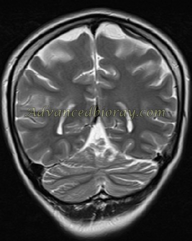
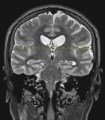
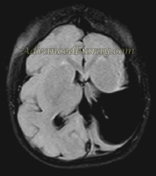
12-year-old right-handed boy, presented with history of complex partial seizure since the age of 18 months; In the conventional MRI, there is focal abnormal signal area located in the right fronto-temporal lobe. There is deformed normal gray matter and represent malformed configuration with thickening and has occupied frontal lobe white matter.
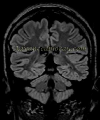
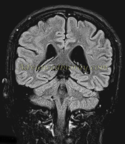
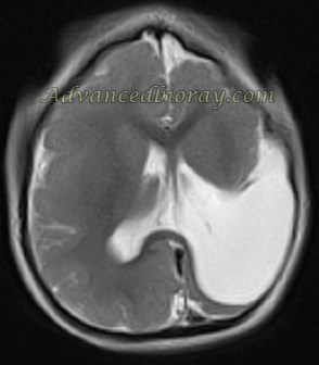
A 12-year-old right-handed girl presented with history of epilepsy since the age of 6 years. MRI represents, focal abnormal signal area located in the left fronto-temporal lobe without radiation band suggests FCD IIA.
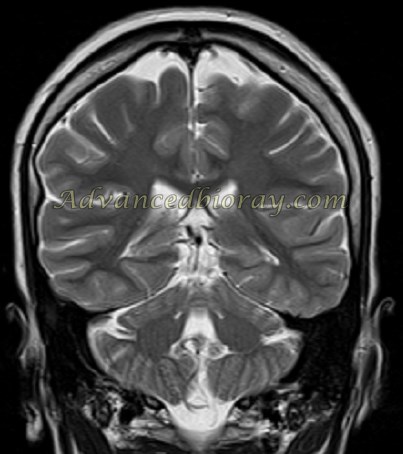
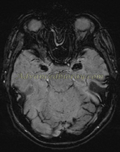
A 13-year-old right-handed girl presented with history of epilepsy since her infancy. MRI represents, wide band of heterotopic gray matter just under cortex. The overlying sulci are too few in number and very shallow; the gyri are abnormally wide. This is compatible with band heterotopia. Final diagnosis was Lissencephaly case with Lennox-Gastaut presentation
A patient with refractory seizure representing open lip Schizencephaly in the left hemisphere, TI and T2 WI MR in the same child shows the abnormally thick and featureless cortex lining the schizencephaly clefts.
A 14-year-old female patient a history of partial seizures and hoarseness when she was 4 years old. She had a brother with some skin scars on his body with a history of recurrent upper respiratory infections. Urbach–Wiethe disease was confirmed