Recent studies have explored the use of Quick Brain MRI (QB-MRI) with diffusion-weighted imaging (DWI) as a first-line imaging modality for diagnosing pediatric stroke. QB-MRI has shown higher sensitivity for detecting ischemia compared to CT scans (100% vs. 27.3%) and can lead to faster diagnosis. It is particularly useful for younger patients and those already hospitalized. A rapid sequence MRI protocol using DWI and fluid-attenuated inversion recovery (FLAIR) has also demonstrated high sensitivity and specificity in identifying ischemic strokes. While MRI is generally more informative than CT in pediatric stroke cases, the choice of imaging modality may depend on factors such as patient age and clinical presentation. These findings suggest that QB-MRI or rapid sequence MRI protocols could be considered first-line imaging studies for children presenting with suspected acute stroke, potentially improving diagnostic accuracy and reducing time to treatment. Some of the samples from our database are presented for researchers in this field.
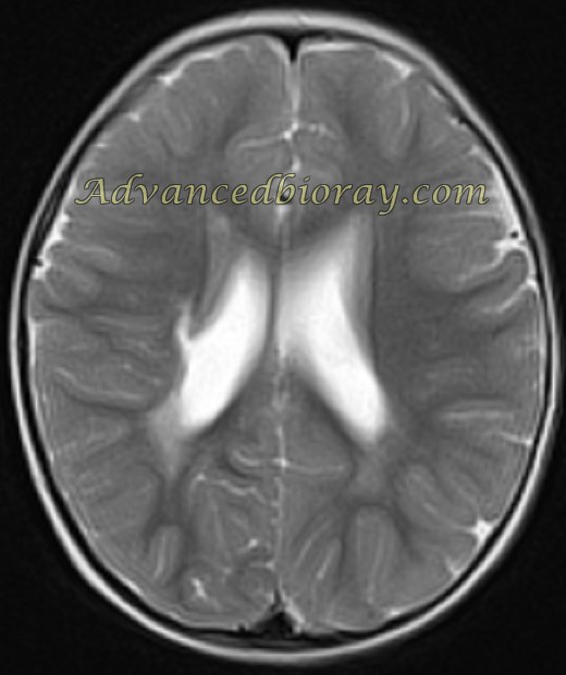
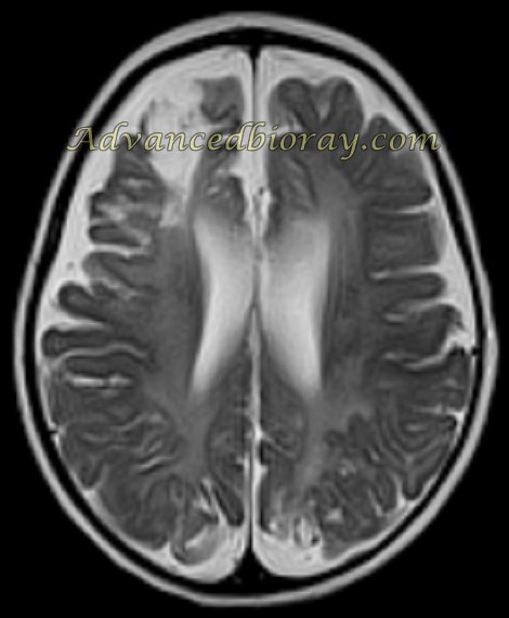
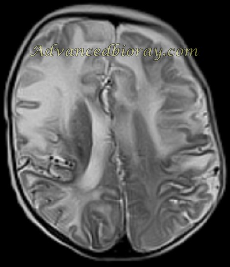
Case No. 1 An 18-month-old girl with chronic hemorrhagic infarction in the right periventricular region, focal malacia, and volume loss.
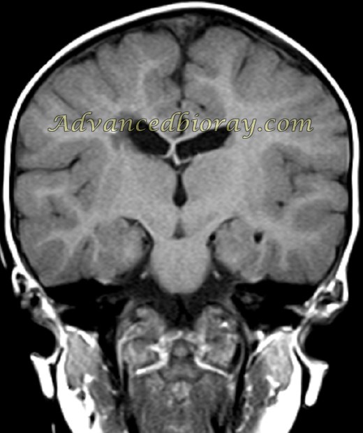
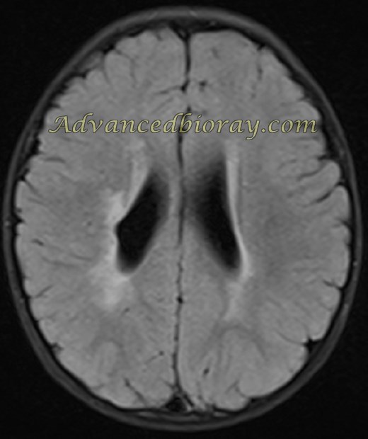
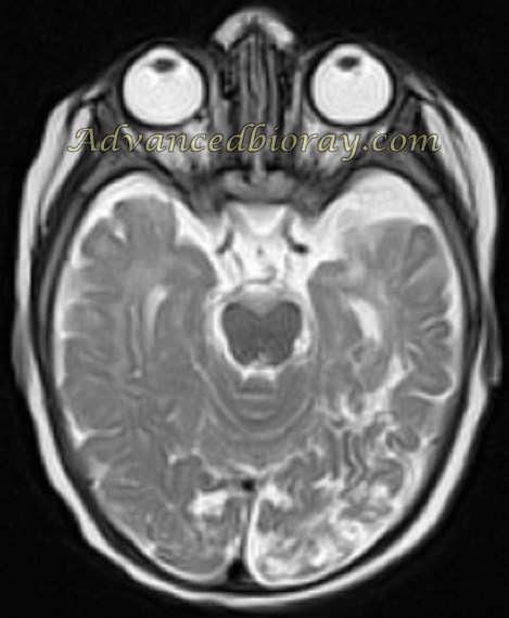
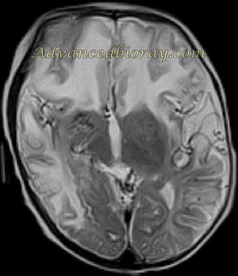
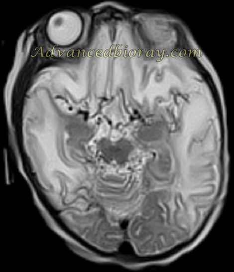
Case No. 2
A 7-month-old infant with several multifocal nonterritorial infarcted zones and focal encephalomalacia. Further evaluations confirmed a case of mitochondrial disorder “MELAS”
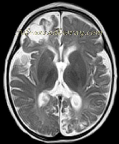
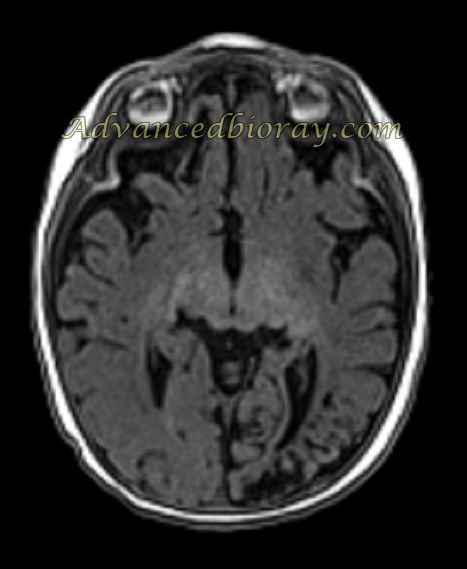
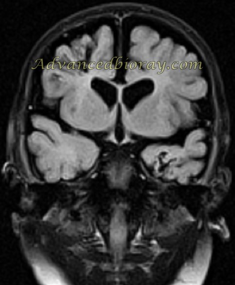
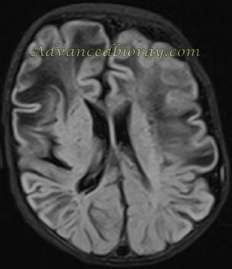
Case No. 3
A 5-month-old boy with MRI of the brain showing prominent collateral circulation in the cortex of both hemispheres with vascular infarctions, diagnosed as Moyamoya disease.