One of the interesting fields in neuroimaging is rare cases not so often seen in daily work.
Rare neurological disorders present diagnostic challenges due to their diverse clinical manifestations making neuroimaging crucial for diagnosis and management. MRI plays a vital role in identifying characteristic features of rare disorders eps in rare syndromes, neurocutaneous disorders, phakomatosis, and neurometabolic disorders. The rare cases and rare presentations of common disorders are introduced on their pages such as neurometabolic disorders but here we introduce other rare cases Even the unusual appearance of disorders may be attractive for teaching points and sharing in clinical practice. We tried to gather our unusual cases during 20 years of focused working in the neuroimaging field and we share some of these here. Archive of rare disorders could be important in diagnosis as well.
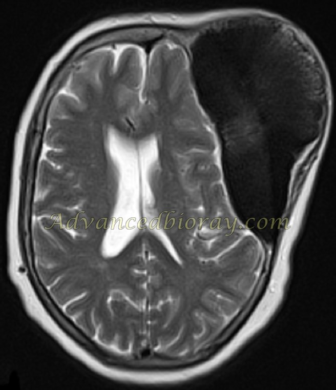
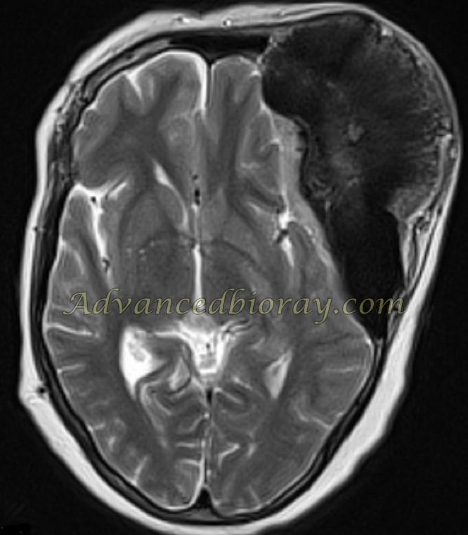
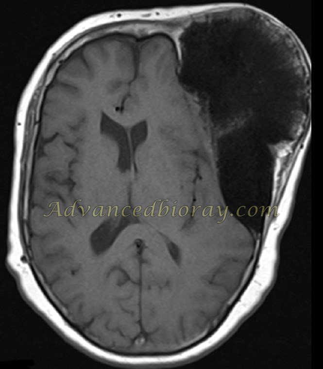
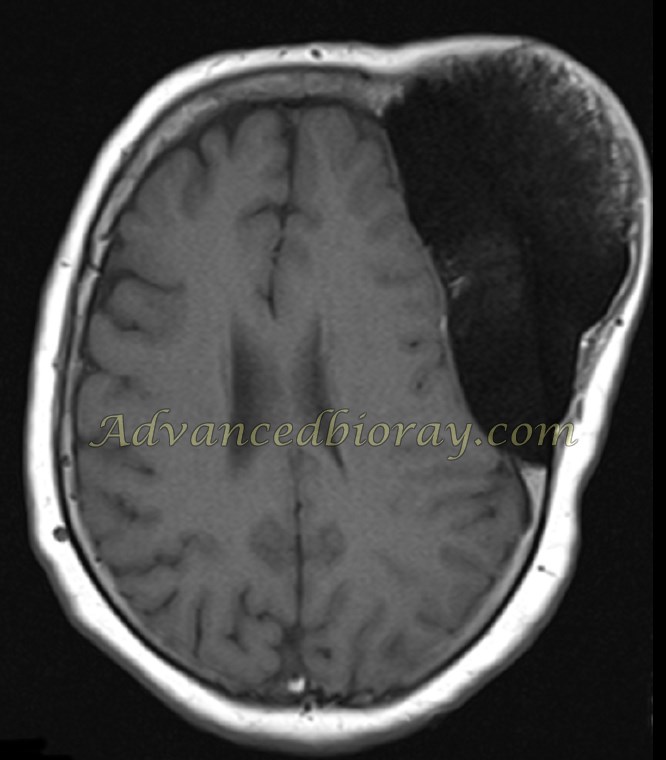
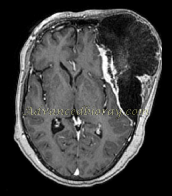
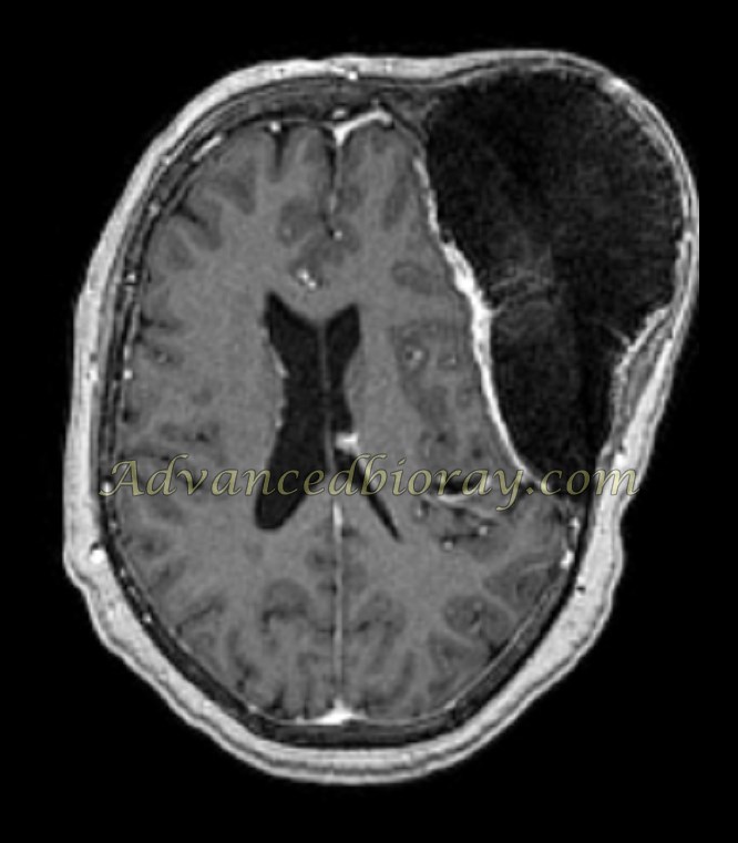
Case No 1
This is a rare presentation of a common tumor in the brain. The patient is a 65-year-old woman with a large mass in the head which was neglected for many years after the MRI presented as a large meningioma with intra-cranial involvement and extension to cranial valut and subcutaneous region.
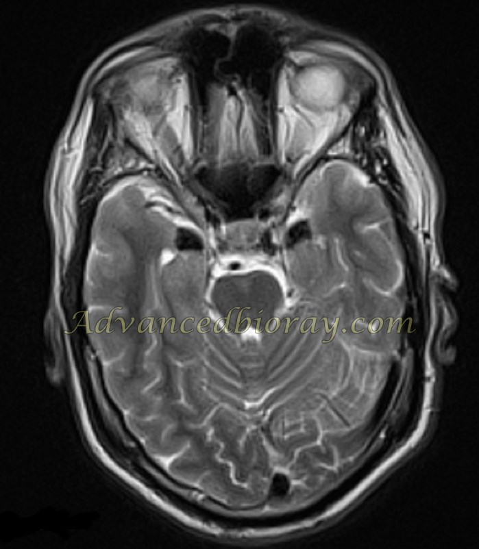
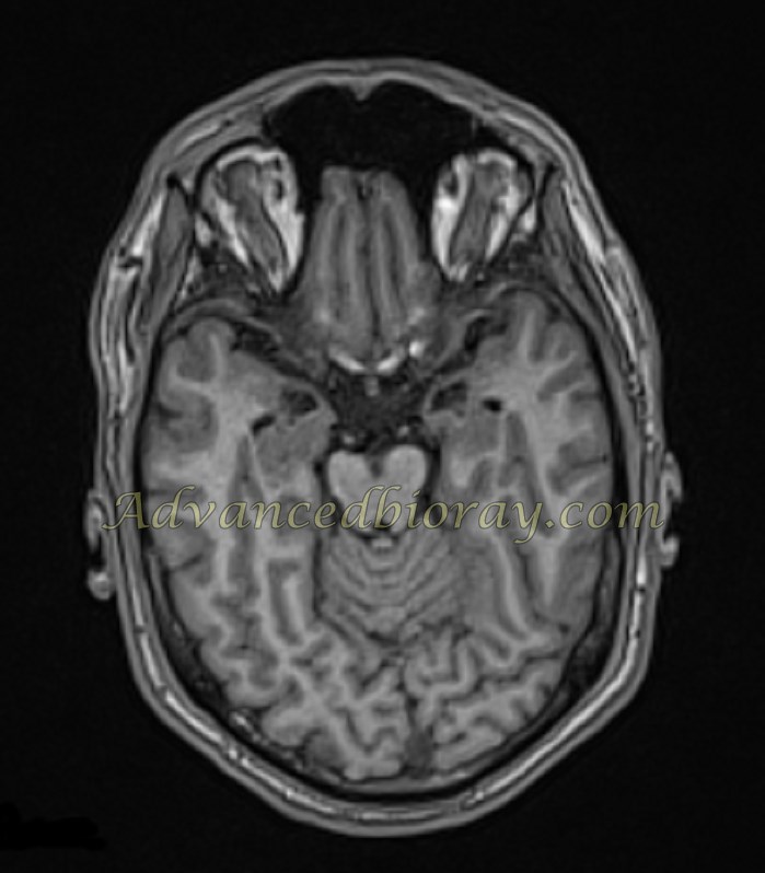
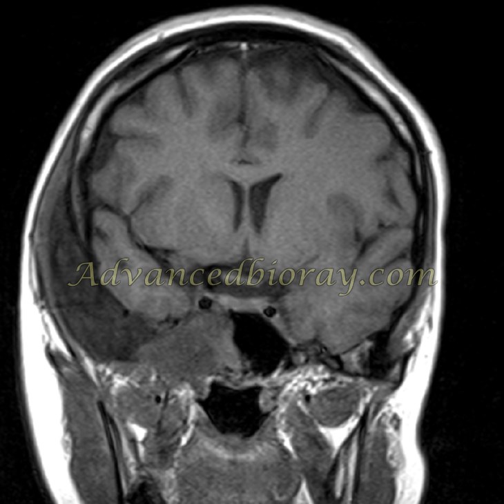
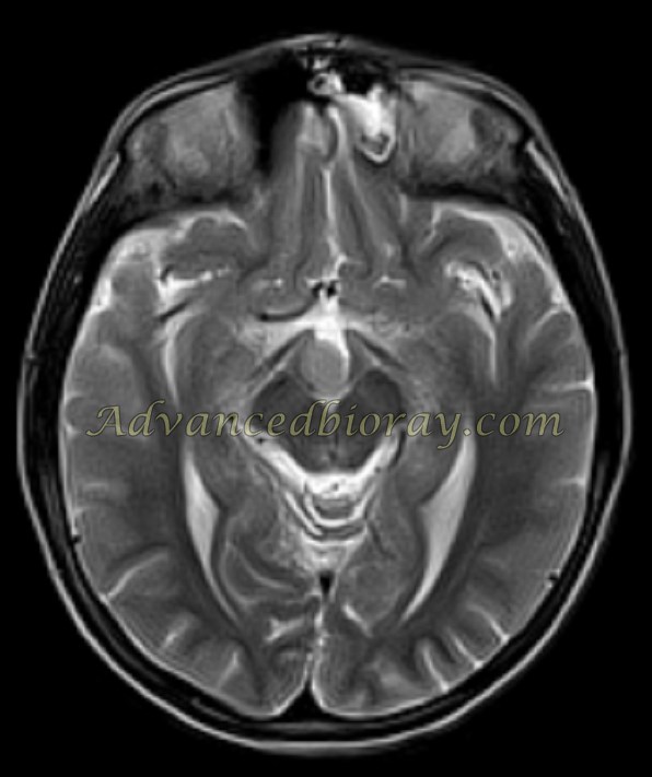
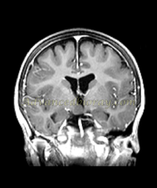
Case No 2
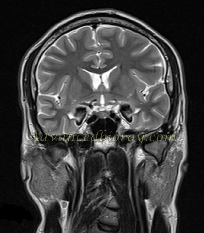
A 19-year-old boy presented with hoarseness and skin disorders with a long history of refractory seizure.
In the obtained MRI symmetrical and bilateral calcification of both Amygdala as a signal void abnormality is the anatomical location of the amygdala is seen. It is a typical case of Urbach-Wiethe disease (UWD).
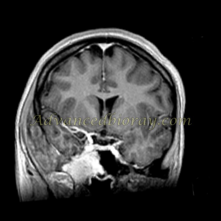
Case No 3
17 year old woman was referred with bulging in the right maxillofacial region, In the MRI with and without contrast media and correlated post-op evaluation the patient had dual lesion :
First: Fibrous dysplasia with involvement of the right orbital walls and temporal bone,
Second: abnormally enhanced meningioma in the right parasellar region with encroachment of the right sinus cavernous.
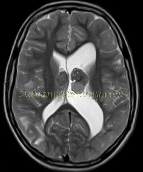
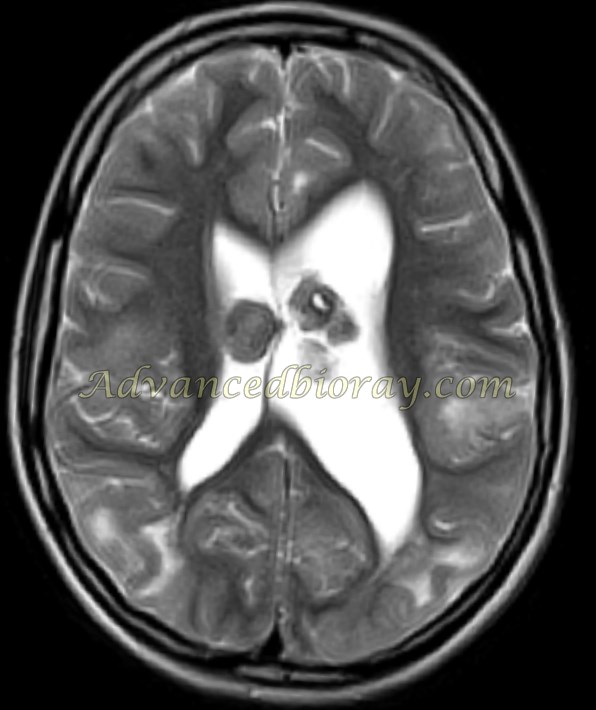
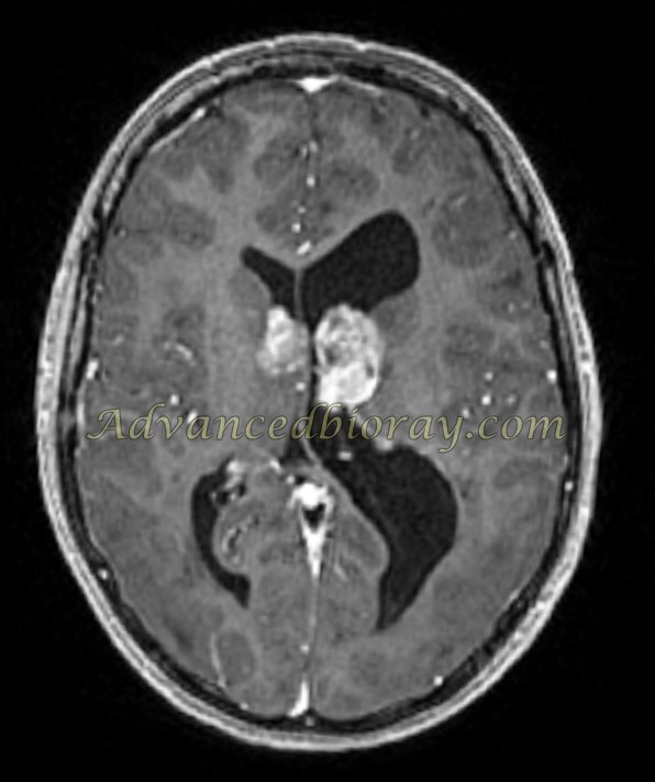
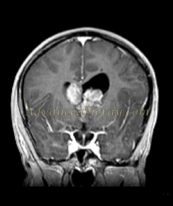
Case No 4
This is a known case of refractory seizure with the recent headache. It is the full presentation of TS (Tuberous Sclerosis ) as several subcortical hamartoma and subependymal calcified nodules. Also, in the post-contrast study, abnormally enhanced tumoral masses in the vicinity of bilateral foramen are compatible with Giant cell astrocytoma (GCA).
Case No 5
9-year-old boy with Gelastic type seizure which represents Hypothalamic Hamartoma in the 3rd ventricular base and unenhanced in post contrast survey.